 Photonic Materials Technology Section (PMTS)
Photonic & Detector Materials Laboratory (PDML)
Photonic Materials Technology Section (PMTS)
Photonic & Detector Materials Laboratory (PDML)
Development of photonic nanomaterials
Photonic nanomaterials are being developed for nonlinear optical (NLO), long persistence luminescence (LPL), and plasmonic based sensor applications. Along with these nanoparticles with desired shape and size, nanocomposite materials are also being developed and characterized to explore different possible applications.
1. Synthesis of lithium niobate nanoparticles and nanocomposites for linear and nonlinear optical applications
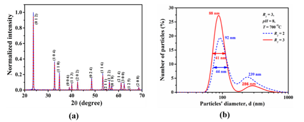
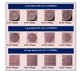
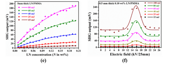
Lithium niobate (LiNbO3; LN) is a technologically important photonic material having ferroelectric, pyroelectric, piezoelectric, electro-optic, acousto-optic, photovoltaic, photorefractive, and nonlinear optical properties with high figure of merit. Recently there has been considerable interest in preparing the LN particles in micron to nano range and utilizing them in different applications where LN bulk single crystal cannot be used. Further, because of several difficulties in the growth and processing of bulk single crystals, there is considerable interest in the preparation of nanocomposite of such materials, which exploit the properties of the nanomaterials, and also offer ease of fabrication and wider functional properties due to the size reduction. This work focuses on developing a new type of nonlinear optical (NLO) material in composite form, consisting of LN nanoparticles (NPs) and polymethyl methacrylate (PMMA) as the host material. LN exhibits various optical and electro-optic properties, and its compositional stoichiometry is crucial for maintaining these properties. The synthesis involved preparing LN NPs (~100 nm) of stoichiometric composition using citrate gel method. These NPs (~100 nm in size) exhibit a bandgap energy of 4.78 eV which is 1 eV higher than reported values for bulk LN single crystals. This elevated bandgap energy facilitates second harmonic generation (SHG) tuning over an expanded spectral range which is required for various applications. PMMA was chosen as the host matrix due to its optical transparency and mechanical properties. A novel synthesis technique was developed to prepare LN/PMMA nanocomposites with oriented LN NPs using an external electric field (EF). Various poled and unpoled nanocomposites were fabricated with different thicknesses and LN NP concentrations to investigate their functionality in the NLO applications. The fabricated LN/PMMA nanocomposites were transparent and optically homogeneous, an essential requirement for their use as optical device elements. The SHG was found to increase as a function of LN concentration for a given fundamental laser pulse energy and thickness. SHG increased upto a certain length of the nanocomposites, beyond which it decreases for a given LN concentration. SHG as a function of poling electric field shows that there is a trade-off between composite thickness and LN concentration to obtain SHG maxima at a specific strength of field for a given pulse energy. The above parametric studies provide crucial insight for fabricating the LN/PMMA nanocomposites to maximize the SHG signal. Additionally, the nonlinear absorption and refraction properties of the nanocomposites were studied, showing that the saturable intensity (IS) and nonlinear refractive index (n2) of LN/PMMA nanocomposites can be tailored by poling the samples during nanocomposite preparation. These nanocomposites have applications in devices required for nonlinear optical applications as well as for ultrafast spectroscopy applications.
|
(a) XRD pattern of Lithium Niobate powders, (b) Lithium Niobate particle size distribution using Dynamic Light Scattering technique |
|
(c) Apparatus & experimental set up for poling of LN/PMMA nanocomposite samples |
|
(d) Unpoled and poled LN/PMMA nanocomposites of different thicknesses |
|
(e) SHG output as a function of LN nanoparticle concentration in LN/PMMA, (f) SHG output as a function of the poling fieldn id="caption"> (Cryst. Growth Des. 8 (2008) 4424-4427; Cryst. Res. Technol. 44 (2009) 305-308; Cryst. Res. Technol. 44 (2009) 1303-1307) |
|
(g) Nonlinear optical properties of the nanocomposites showing saturable intensity (IS) and nonlinear refractive index (n2) of LN/PMMA nanocomposites
[Applied Physics A 126 (2020) 611 (1-8); Applied Physics B 127 (2021) 29 (1-10); Bull. Mater. Sci. (2024) 47:77 (1-11)] |
2. Synthesis of NIR emitting Cr3+ doped zinc gallogermanate (Zn3Ga2Ge2O10:Cr3+) nanoparticles for bioimaging applications
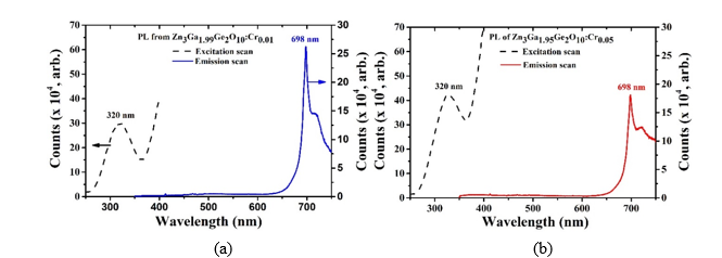
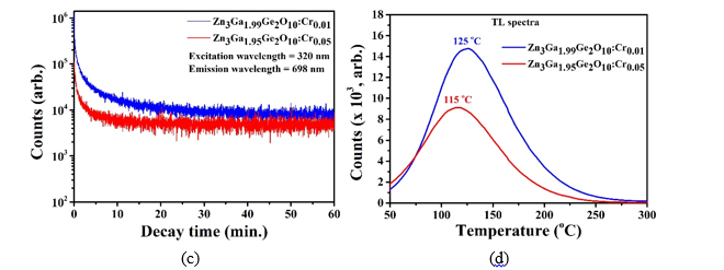
Optical imaging has attracted a lot of interest in recent years, because of its benefits of high sensitivity, reduced cost and time effectiveness, portability, no harmful radiation, and viability for clinical investigations. Strong tissue autofluorescence, light scattering, and phototoxicity are produced when traditional fluorescent probes are excited in situ during in vivo and in vitro imaging. Luminnescent nanoparticles have lasting afterglow that enables optical excitation prior to bioimaging, as well as detection and imaging without external illumination. A type of luminescent nanoparticle namely Chromium doped Zinc gallogermanate (Zn3Ga2Ge2O10: Cr3+) nanoparticles were successfully synthesized using citrate gel method. The average particle size was below 50 nm which is required for cellular uptake. The particles exhibit strong long persistence luminescence at NIR (698 nm) when excited with UV (320 nm) light (Fig.(a) & (b)). Through kinetic scan (Fig. (c)), it was confirmed that the nanoparticles were behaving as long persistent luminescent (LPL) materials. The trap depth of electron was estimated from TL-glow curve (Fig. (d)), which are 0.7963 eV and 0.7763 eV respectively for Zn3Ga1.99Ge2O10:Cr0.01 and Zn3Ga1.95Ge2O10:Cr0.05 nanoparticles respectively. Fluoresce-based bioimaging experiments on HaCat cells under in situ excitation of 254 nm with emission at 698 nm confirmed that the Cr-doped ZGGO nanoparticle can be used as a probe in bioimaging (Fig (e, f)).
|
((a) The PL spectra obtained from emission and excitation scan in case of Zn3Ga1.99Ge2O10:Cr0.01, (b) the PL spectra obtained from emission and excitation scan in case of Zn3Ga1.95Ge2O10:Cr0.05 |
|
(c) The kinetic scan for PL at 698 nm wavelength for both the samples and (d) TL spectra obtained for both the samples |
|
e) Bright field and (f) fluorescence image of HaCat cells using Zn3Ga1.99Ge2O10:Cr0.01 nanoparticles.
[Proc. NLS-32 (2024) RRCAT Indore] |
3. Synthesis of zinc oxide nanorods on gold nanoparticles coated glass substrate for LSPR based sensor applications
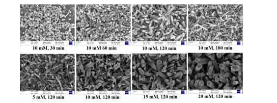
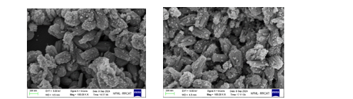
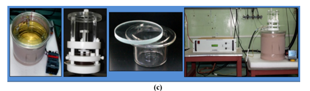
A three-dimensional (3-D) nanostructured sensor surface has been developed for Localized Surface Plasmon Resonance (LSPR)-based sensing applications with the help of a wet chemical synthesis process through the optimization of different synthesis parameters. These sensor surfaces were fabricated using gold nanoparticles (AuNPs) and Zinc Oxide nanorods (ZnO NRs) for enhancing the sensitivity of plasmonic biosensor. The ZnO NRs were synthesized using a hydrothermal method on a ZnO-seed layer applied to the AuNPs-coated glass substrate. The ZnO seed particles, which act as nucleation sites for ZnO NRs growth, were synthesized using the sol-gel method. The ZnO seed layer was coated on the AuNPs-coated glass substrate using drop casting technique. The seeded AuNPs were synthesized by citrate reduction method and which was coated on glass for adhesion between glass surface and ZnO seed particles. The AuNPs were immobilized on the glass substrate with the adhesion of APTES (3-Aminopropyltriethoxysilane) layer. The average length and diameter of ZnO nanorods were controlled by varying the concentration of growth solution and growth time (Fig.(a)). The synthesized ZnO NRs were then decorated with gold nanoparticles of diameter ~ 20 nm -30 nm (Fig. (b)) for plasmonic applications.
|
(a) SEM images of synthesized ZnO nanorods for different solutions and growth time |
|
(b) SEM images of synthesized ZnO nanorods coated with gold nanoparticles |


![(g) Nonlinear optical properties of the nanocomposites showing saturable intensity (IS) and nonlinear refractive index (n2) of LN/PMMA nanocomposites
[Applied Physics A 126 (2020) 611 (1-8); Applied Physics B 127 (2021) 29 (1-10); Bull. Mater. Sci. (2024) 47:77 (1-11)]](photonic_5.png)
![(e) Bright field and (f) fluorescence image of HaCat cells using Zn3Ga1.99Ge2O10:Cr0.01 nanoparticles. [Proc. NLS-32 (2024) RRCAT Indore]](photonic_8.png)