|
|
Role of radiative decay of valence plasmons in transmission spectra of Si, SiNx and PET membranes
[Solid State Communications 156 (2013) 12-15]
Plasmons in metallic samples are excited by intraband as well as interband transitions of electrons. In case of non-metallic samples, they are excited by interband transitions only. In the present study, plasmons generated by inter- band transitions of electrons are denoted by valence plasmons (VPs). Valence plasmons are different from volume plasmons which are excited by intra- band transitions of free electron gas. Interband transitions can take place when energetic particles (photons, electrons, protons and ions) deposit an sufficient amount of energy into a material, as a result photoelectrons, excitons along with valence bulk plasmons (VBPs) and valence surface plasmons (VSPs) are generated in materials (metals, semiconductors and insulators)
When high energy photons, electrons and ions pass through a material, they suffer quantum energy losses in the range 5-40eV. These losses are explained on the basis of collective oscillations of VPs. In PES, the photoelectrons lose their kinetic energy by inelastic scattering with VPs and hence, plasmons peaks are observed at higher binding energy (BE) with respect to the BE of core photoelectrons. The query is about possible decay mechanisms of VPs. Probable decay processes of VPs are (1) by emission of secondary photoelectrons of energy equal to the difference of VPs energy and work function of the material, (2) by generation of electron - hole (e-h) pairs inside the material and (3) scattering with electrons and phonons. These (e-h) pairs will decay either radiatively or non-radiatively. It is expected that the decay of VPs into e-h pairs and subsequent radiative recombination should increase transmitted and reflected intensities at VPs energies. On the other hand, non-radiative decay of plasmons will result in minima in transmitted and reflected intensities.
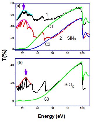
In the present work, transmission spectra of Si, SiNx and polyethylene terephthalate (PET) membranes are measured in vacuum ultra violet (VUV) energy range. Maxima in the transmitted intensity are observed at VPs energies of Si, SiNx and PET and these maxima are explained by considering radiative decay of VPs.
Present experimental results show that the coupling of photons with valence plasmons is possible. This is first experimental observation of effect of radiative decay VBPs and VSPs in transmission spectra in VUV range of Si, SiNx and PET membranes
|
Figure . (a) Curves 1 and 2 show measured transmission spectra of 50 nm and 100 nm thick SiNx membranes respectively and curves C1 and C2 show corresponding calculated transmission. (b) Transmission spectrum of Si membrane and curve C3 shows calculated transmission. Peaks observed due to radiative decay of valence plsmon in transmission spectra are marked by arrows. |
|
Thermal stability studies of ion beam sputter deposited C/B4C x-ray multilayer mirror.
[Thin Solid Films, 527, 244-249 (2013)]
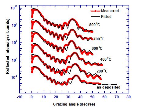
Annealing studies of C/B4C multilayer structure were carried out to understand its thermal stability and subsequent reflectivity performance at the desired wavelengths in the soft x-ray region. Soft x-ray reflectivity technique was employed to analyze the structure of as-deposited and vacuum annealed C/B4C multilayer films have . The results show that the multilayer structure is stable even after 700 °C annealing. Raman spectroscopy indicates graphitization of carbon layer with increasing annealing temperature. Graphitization of carbon results in increases of layer thickness and decrease in density as the same is also observed by soft x-ray reflectivity. The reduction in measured soft x-ray reflectivity at 6.56 and 4.39 nm wavelengths after 800 °C annealing was observed.
|
Figure: (Solid line and filled circle) Soft x-ray angle dependence reflectivity of C/B4C multilayer sample measured at various annealing temperature near Boron K edge (6.56 nm) and their best fits are shown by solid line respectively. |
|
Stability and normal incidence reflectivity of W/B4C multilayer mirror near the boron K absorption edge.
Appl. Opt. 52, 6126-6130 (2013).
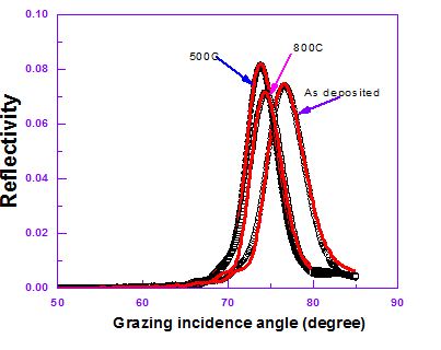
Multilayer structure consists of alternating layers of W and B4C has been deposited using magnetron sputtering system. The structure of as-deposited and vacuum annealed W/B4C multilayer sample has been characterized using grazing incidence x-ray reflectivity, grazing incidence diffraction and the normal incidence reflectivity has been measured using synchrotron radiation. A two layer model consists of tungsten and boron carbide is presented. The multilayer structure is found stable after 800 °C annealing. Grazing incidence x-ray diffraction measurements suggested that W is polycrystalline with small grain size. No signature of tungsten carbide or tungsten boride formation could be observed during the annealing treatments. A near normal incidence soft x-ray reflectivity of ~8.3% was obtained at 6.8 nm wavelength.
|
Figure: Measured and fitted SXRR data of as-deposited and annealed sample at 500 °C and 800 °C at 6.7 nm wavelength. The open circle, filled circle and open diamond represent the measured SXRR data of as-deposited sample, annealed at 500 °C and 800 °C respectively. |
|
Cleaning of Carbon Layer from the Gold Films Using a Pulsed Nd:YAG Laser
Applied Surface Science 283 (2013) 612- 616
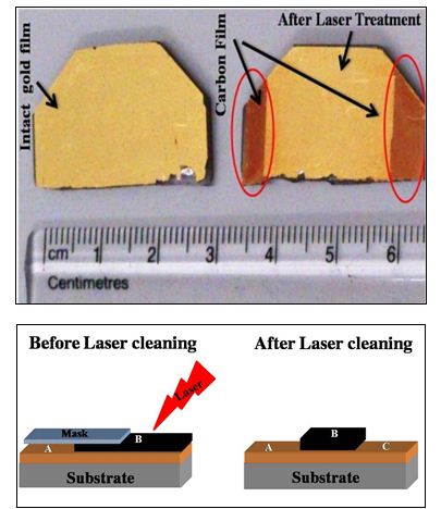
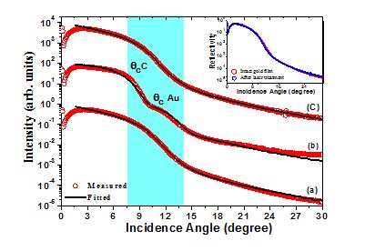
Hydrocarbon cracking and carbon contamination of optical elements in soft x-ray spectrometers and synchrotron radiation beamlines is a severe problem. Carbon contamination seriously affects the optics performance. In the present work, an Nd:YAG laser providing 2 mJ of pulse energy and 100 ns of pulse duration has been used for carbon cleaning experiments. The laser cleaning is a non-contact, accurate, efficient and safe process. A surface area of 48 cm2 having ~20 nm thick carbon layer on gold surface has been removed with six number of laser passes and with 80% laser spot overlapping without any change in surface roughness of the underneath gold film. Cleaning effect and subsequent film quality after laser irradiation has been analyzed using X-ray photoelectron spectroscopy, soft x-ray reflectivity techniques. Atomic force microscopy was used to analyze surface morphology before and after laser cleaning process. Power spectral density function was calculated over large frequency range of 10-1 to 10-4 nm-1 to understand topographic data.
|
Fig.: Photograph and schematic diagram of the sample used for laser cleaning experiments are shown : (A)intact gold film, (B) carbon coated gold film, (C) carbon removed gold film after laser treatment |
| |
|
Fig.: Soft x-ray reflectivity measurements carried out before and after laser cleaning experiments suggest that the reflectivity pattern obtained after the carbon removal are in agreement with that of intact gold film. This indicates that the carbon has been successfully removed (see inset). a) reflectivity pattern of intact gold film, b) carbon coated gold film and c) gold film after carbon removal. |
|
Study on effective cleaning of gold layer from fused silica mirrors using nanosecond-pulsed
Nd:YAG laser. [Applied Optics, 52, 7540 (2013)
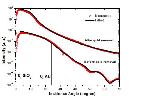
A study on effective laser cleaning of gold layer deposited on fused silica substrates has been carried out using nanosecond-pulsed Nd:YAG laser. The influence of pulse duration, beam incidence angle, spot overlapping, laser fluence, and number of passes on cleaning efficiency has been investigated. An approximately 48 nm thick gold layer from a surface area of ~48 cm2 has been cleaned in 3 min. Laser clean quality and efficiency has been analyzed using microscope, scanning electron microscope (SEM), and angle-dependent reflectivity measurement techniques using SR beamline. Optimization of cleaning parameters resulted in a cleaning efficiency of ~98%. This study provides an alternate and low-cost solution for reuse of gold-coated, damaged mirrors.
|
Fig.: Soft x-ray reflectivity measurements of gold coated fused silica and gold removed fused silica substrate suggest that the laser technique can be used to remove gold layer from fused silica substrate effectively. |
|
NbC/Si multilayer mirror for next generation EUV light sources
In this study we report a new multilayer combination comprised of refracting layers of niobium carbide and spacer layers of silicon as a more stable and high reflecting combination for the 10 - 20 nm wavelength region. The reflectivity of the new combination is comparable to Mo/Si conventional mirrors. Annealing experiments carried out with NbC/Si multilayer at 600°C temperature showed a ~2.5% drop in the soft x-ray reflectivity along with a marginal contraction in the multilayer period length. The multilayer structure is found stable after the heat treatment. Crystallization of the niobium carbide and silicon layers is responsible for the compaction in the period length as revealed by the grazing incidence x-ray diffraction measurements. No signature of silicide formation or any other chemical species could be detected. The multilayer structures were grown by ion beam sputtering technique using a compound target of niobium carbide. Soft x-ray reflectivity measurements performed at the Indus-1 and BESSY-II synchrotron radiation sources are found in good agreement with the simulations.
|
 Calculated soft x-ray reflectivity profile of Mo/Si and NbC/Si multilayers with identical structural parameters (d= 6.3 nm,Γ = 0.428, σ=0.3 nm, N= 51 layer pairs). At 85.0° incidence angle, the peak reflectivity of two multilayers is slightly different by ~2.5 %.
|
Quantitative determination of higher harmonic contents in the soft x-ray spectra of toroidal grating monochromator using a reflection multilayer
Grating monochromator causes a problem of higher harmonic contaminations in a synchrotron beamline operating in the soft x-ray/ vacuum ultra violet region. Generally gratings are used to experimentally determine the higher harmonic contaminations. In this method, the relative contribution of contaminant wavelengths is measured with respect to the first harmonic wavelength (desired wavelength). The quantitative fit of grating spectra is rather complex and therefore qualitative analysis is carried out.
Analysis of multilayer reflectivity data has become rather simple with recent advances in the theoretical modeling. Therefore we propose to use a multilayer mirror and analyze its reflectivity data for quantitative determination of harmonic contamination in a soft x-ray beamline. In the present study we used a Mo/Si multilayer of d = 97 Å to quantify the spectral purity of 600 l/mm toroidal grating at the reflectivity beamline of Indus-1 450 MeV synchrotron source. The measured reflectivity spectra at each wavelength is analysed and actual contribution of higher harmonics in the incident beam is obtained.
|
 Measured reflectivity curve at λ= 260 Å is fitted by taking into account the fractional contribution of higher harmonics λ/2 = 130 Å (dotted curve) and λ/3 = 86.66 Å (dashed curve).
|
|
Probing porosity at buried interfaces using soft x-ray resonant reflectivity
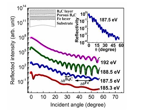
The optical constants of electron beam evaporated boron carbide are measured near boron K-absorption edge. Near the edge, the dispersion part of refractive index shows a sign reversal. Simulated reflectivity profiles near the absorption edge of boron are used to show the utility of soft x-ray resonant reflectivity as a sensitive tool for probing selected buried interfaces. This is due to high and tunable scattering contrast. The simulated resonant reflectivity profiles are sensitive to porosity and position of the porous layer containing the resonating atom. This is experimentally demonstrated through soft x-ray resonant reflectivity measurements of B4C-on-Fe bilayer structure
|
Fig.: Soft x-ray reflectivity profiles of B4C-on-Fe bilayer film near boron K-absorption edge. [J.Appl.Phys. 107, 023529 (2010)].
|
|
|
Study of polished zinc sulphide surface
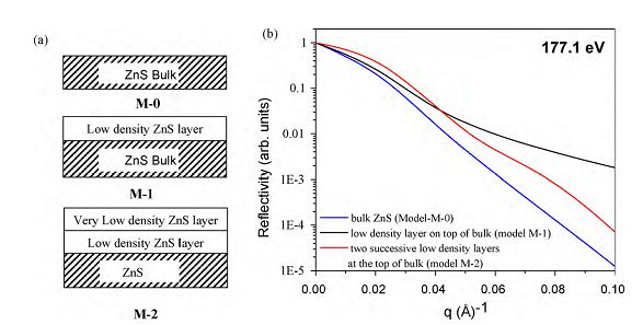
Resonant soft x-ray reflectivity measurements at and near the L absorption edge of sulphur have been performed on mechanically polished zinc sulphide using reflectivity beamline. A sulphur rich surface (∼15nm thick) consisting of two layers with gradient electron density distribution was uniquely determined. As compared to bulk ZnS, the top layer has ∼30–50% less electron density whereas, the intermediate layer has ∼10–18% less electron density. Conventional hard X-ray reflectivity measurement at Cu Kα wavelength also indicates low electron density (sulphur rich) surface of ZnS but the technique was found insensitive for unique determination of electron density distribution. Optical constants of ZnS in the soft x-ray region (100–250 eV) have been reported for the first time and were in good agreement with the theoretically reported values.
|
Fig.: The figure shows that the soft x-ray reflectivity near Sulphur L- edge gives a significant contrast corresponding to change in surface density in ZnS sample [Applied Surface Science 257 (2010) 210].
|
|
|
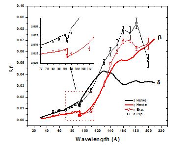
Optical properties of Indium Phosphide in 50-200Å; wavelength region using reflectivity technique
The optical constants of Indium Phosphide (InP) in the soft x-ray region of 50-200Å are determined from the angle dependent reflectivity measurements. The derived optical constants are compared with tabulated values of Henke et al. Experimental values of δ and β are in close agreement with the tabulated values in lower wavelength region of 50-120Å. The experimental value indicates an edge shift of 0.4Å towards lower wavelength side from the phosphorous L-edge value of 92Å. However, above 120Å region where Indium N2 edge falls at 160.7Å there is huge difference between experimental and tabulated values are observed. Both delta and beta values are significantly higher. In contrary to tabulated values of β/δ ratio which is more than one above 140Å region, the experimental measured ratio is found to be less than one. This study presents first reported experimental values of optical constants for InP in this wavelength range.
|
Fig.: Measured optical constants of InP material in 50-200 Å wavelength region [Appl. Opt. 49, 5378 (2010).]
|
|
|
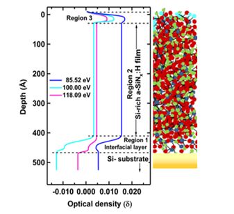
Growth kinetics and compositional analysis of silicon rich a-SiNx:H film: A soft X-ray reflectivity study
In this study, optical index profile derived over extended energy region of soft x-ray regime combined with soft x-ray reflectivity technique has been utilized to obtain depth graded compositional details and growth kinetics. This method has been applied near the Si L2,3 edge for non destructive characterization of Si-rich silicon nitride (SRSN ) film to reveal buried Si-rich interfacial layer. Further, the combined study of soft x-ray reflectivity and optical density obtained from the reflectivity fitting at various photon energies provide a qualitative estimation of the film composition and its growth. The new method suggested in this work might be capable in improving the understanding of growth kinetics in other materials also.
|
Optical density profiles across the depth of SRSN film as derived from SXR fit. Appl. Phys. Let. 97 (2010) In press. |
|
|
Study of optical response near the absorption edge
Fine structure features of energy dependent atomic scattering factor near the atomic absorption edge, is used for characterization of low Z containing hard-condensed matter thin films. Near the atomic absorption edge, reflectivity
Journal
Study of optical response near the absorption edge using vacuum ultra violet/soft x-ray reflectivity beamlines on Indus-1,
G S Lodha,M Nayak,M H Modi,A K Sinha,R V Nandedkar,
Journal of Physics: Conference Series 80 (2007) 012031
|
|
Smoothening of tungsten�carbon interfaces and change in interface asymmetry on heat treatment
|
 |
 |
interface modifications in tungsten carbon multilayer are analyzed by performing isochronal annealing experiments in the range of 200-800 -C. X-ray reflectivity data revealed that the roughnesses are increasing in the W/C multilayer on going from bottom to top layer. The two interfaces viz W-on-C and C-on-W show an asymmetric change in the roughness values. Transmission electron microscopy results indicate that the interfaces are morphologically smooth but are chemically diffused in nature. Roughnesses are smoothening out after annealing at 400 -C, which results in an increase in the multilayer reflectivity
|
Soft x-ray resonant reflectivity for determination of interlayer
Near the absorption edge, fine structure features of energy-dependent atomic scattering factor are sensitive to the composition, and can be exploited for determination of composition at the buried interfaces. This technique is demonstrated for a Mo-Si multilayer system using soft x-ray resonant reflectivity measurement
|
|
|
|
Network compaction and surface deformation in the hydrogenated silicon nitride film
Modifications in a hydrogenated silicon nitride film by soft x-ray/VUV (vacuum ultra violet) radiations are investigated using in situ soft x-ray reflectivity measurements at Indus-1 synchrotron source. The illumination experiments are performed at 10° grazing incidence angle at which the majority of incident radiation (belonging to 10 eV-250 eV) are restricted to ~8.0 nm depth, except near the Si-L absorption edge (100 eV) where changes. Resultantly, the film density has increased along with a change in surface morphology. Due to illuminations, hydrogen bonds responsible for voids and network deformation are more likely to break and pave the path for the formation of more compact Si3N4 network. Evolution of hydrogen changes the surface morphology significantly. Atomic force microscopy confirms the formation of nano clusters at the surface. The out diffusion of hydrogen near the surface is responsible for surface deformation.
|