|
Beamline overview
Material investigations under extreme conditions (high pressure/ temperature) provide vital information about microscopic interactions through structural evolution and/or realization of new phases which may have unusual properties. To serve this purpose, the in-situ structural determinations under extreme conditions are primarily performed using x-ray diffraction (XRD), a versatile non-destructive technique. There are two main variants of x-ray diffraction technique viz. angle dispersive x-ray diffraction (ADXRD) and energy dispersive x-ray diffraction (EDXRD). Both the techniques have their own merits. EDXRD technique is advantageous in case of constrained geometries and kinetics investigations whereas ADXRD provides high resolution diffraction data for detailed structural refinement. An ED/AD-XRD beam line, incorporating both these variants and especially optimized for material investigations under extreme conditions has been developed and commissioned at port no. BL-11. This beam line is being routinely used by several national and international users.
Beamline specifications
| |
EDXRD |
ADXRD |
| SR source: |
Bending Magnet 1.5T |
Bending Magnet 1.5T |
| Energy range: |
10-70 KeV @ 2.5 GeV, 300 mA |
Monochromatic tunable
10 Kev to 30 KeV
|
| Angular range for diffraction |
±25° |
Depends on sample to detector distance, typically ± 45 °
|
| Q Range: |
1.3 to 15 Å-1 (for 2θ =25° ) |
~Up to 12 Å-1 at 30 keV
|
| Detector resolution |
145 eV at 5.9 KeV; 475 eV at 122 KeV |
Mar345 area detetctor with 0.1 * 0.1 mm2 pixel
|
| Resolution |
2% (Geometrical) |
4.7 * 10-4 @ 17 KeV (calculated)
|
| Spot size at sample |
~100*100 μm (which can be increased up to 5*5 mm) |
~ 50x30 μm (using compound refractive lens)
|
| Total Flux at sample |
1011 photons/s (for 300 mA @ 2.5 GeV and 100*100 μm spot size) |
1010 photons/s (for 300 mA @ 2.5 GeV and 1x1 mm2 spot size)
|
Experimental setup
The front end for the Extreme conditions ED/AD-XRD beamline delivers a 1 mrad white beam. This beam is transported to the experimental station through various components viz. beam stopper, primary slit and precision slit. Primary slit and precision slit cut down the size of synchrotron radiation (SR) to 100μm * 100μm at the sample. There are two variants at the experimental station viz. the EDXRD variant and the ADXRD variant.
1) EDXRD variant
A custom made 8-axis sample stage goniometer from Huber is installed for sample mounting. This goniometer has maneuverability of XYZ translation, rotation and tilt. The data acquisition system comprises of a high resolution HPGe detector mounted on a detector arm attached to the 2θ stage of the goniometer. The detector is preceded by a cleaning slit and a point slit. Diffraction angle is defined by cleaning slit and point slit within the precision of ~0.001°. Cleaning slit also helps in reducing the background arising from the Compton scattered x-rays. Main features of EDXRD variant are tabulated below.
8 axis motorized goniometric stage |
Bottom XY- stage ; Θ-2Θ stage; Sample XYZΧ stage |
2Θ detector arm with travel range ±25° |
Detector: High Purity Ge detector |
 |
Fig. EDXRD variant at BL-11 |
2) ADXRD variant
To complement the utilization of EDXRD beamline, it has been adapted for ADXRD measurements. This has been achieved by monochromatizing white synchrotron beam using Si (111) channel cut monochromator. This adaptation has been ergonomically conceived using the existing infrastructure at the beam line and switching between the two variants is easily achieved. In order to avoid diffraction peaks from gasket material used for high pressure measurements, a compound refractive lens (CRL) based micro-focusing arrangement has also been incorporated at the beamline. These CRL's have been indigenously developed at x-ray lithography (BL-7) of INDUS-2 synchrotron source.
 |
Fig. Monochromatization using single crystal Si (111) channel cut monochromator
|

|
Fig. Micro-focusing using indigenously developed Compound refractive lens (CRL)
|

|
Fig. Diffraction image collection at MAR345 imaging plate
|
Beamline capabilities
- Energy dispersive x-ray diffraction measurements
- High pressure powder diffraction (Up to Megabar)
- High temperature powder diffraction (inside capillary up to 1500° K)
- Energy dispersive grazing incidence x-ray diffraction
- Ambient and High temperature (up to 700°K)
- Angle dispersive x-ray diffraction
- High pressure powder diffraction (Up to Megabar)
- High pressure single crystal diffraction
- High temperature powder diffraction (inside capillary up to 1500° K)
- Total x-ray scattering measurements for pair distribution function analysis
 |
Fig. Grazing incidence EDX adaptation for thin films
|
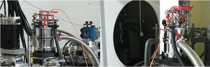 |
Fig. Adaptation of ED/ADXRD setup for high temperature measurement up to 1500° K
|
 |
Fig. Off-line ruby pressure measurement setup
|
A few recent studies performed using BL-11
-
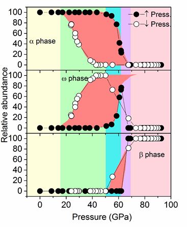
High pressure study on α⇔ω⇔β transition sequence in several pure and impure samples of Hf metal
Journal of Applied Physics, 115, 233513 (2014)
|
Fig. Evolution of volume fraction of alpha, omega and beta phases of Hf as a function of pressures
|
-
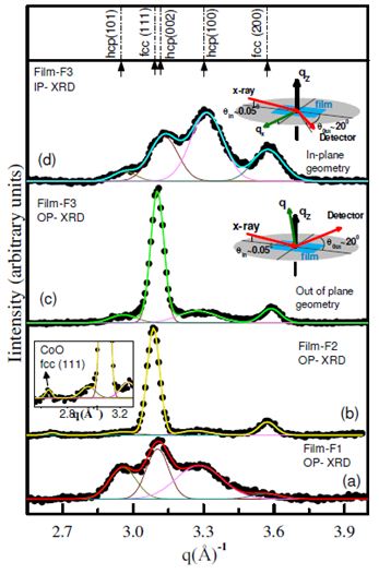
Ex-situ in-plane and out-of-plane diffraction measurements revealing the growth of Co on oxidized interface with preferential orientation of c-axis perpendicular to the film plane
Journal of Physics D: Applied Physics, 47, 105002 (2014)
|
Fig. Grazing incidence x-ray diffraction of Co/CoO/Co thin film
|
-
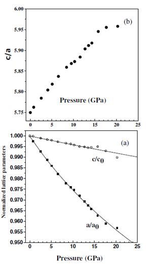
Stability of ambient rhombohedral structure of CuCrO2 upto ~ 23 GPa however with large anisotropy in axial compression with c-axis compressibility, kc = 1.26 * 10-3 GPa-1 and a-axis compressibility, ka = 8.9 * 10-3 GPa-1
Journal of Applied Physics, 116, 133514 (2014).
)
|
Fig. Evolution of lattice parameters of CuCrO2 as a function of pressure.
|
-
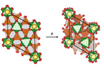
X-ray diffraction studies confirm two phase transformations in NaZr2(PO4)3 around 5 and 6.6 GPa conjectured through Raman scattering measurements
Journal of Solid State Chemistry, 221,285-290 (2015).
)
|
Fig. Structural transition in NaZr2(PO4)3
|
|
Fig. Equation of state of different phases of NaZr2(PO4)3
|
Publications related to Bl-11
2026
- Probing nonlinear structure-orbital correlation for PrOFe 0:9 Co 0:1 as under pressure.,
Debdutta Lahiri, K. K. Pandey, P. Modak, Nitya Ramanan, Mateusz Czyzycki, Gerald Falkenberg, Jai Prakash, Gohil S. Thakur, A. K. Ganguli, Nandini Garg.,
International Journal of Modern Physics B 40 (2026) 2650072.
- Pressure induced order-disorder transformation in Y2Ge2O7: Role of geometric constraints.,
Rahul Kaiwart, Abhilash Dwivedi, K.K. Pandey, Adish Tyagi, Sandeep Nigam, H.K. Poswal.,
Journal of Alloys and Compounds 1058 (2026) 186918.
- Bi3+ Ion as a Performance Curator in a Y2Ge2O7 (Yb-Er) Spectral Convertor: Proliferation of Sensitization Range and Competence.,
Dibya Kanti Mal, Adish Tyagi, Sandeep Nigam, Rahul Kaiwart, Sandeep K. Agarwalla, Himanshu K. Poswal, Vasanthakumaran Sudarsan, Chiranjib Majumder, and Avesh K. Tyagi.,
Inorg. Chem. 2026, 65, 2997−3009.
- Structural analysis of holmium alumino silicate (HAS: Ho2O3–Al2O3–SiO2) glasses using Ab initio molecular dynamics simulation.,
Anurup Das, A.S. Patil, M. Goswami, Velaga Srihari.,
Materials Chemistry and Physics 358 (2026) 132424
2025
- High pressure investigation of structural and electronic behavior of β-ZnMoO4.,
Siddhi Garg, Ashok K Verma, HK Poswal, TCN Nicacio, Daniel Errandonea, Nandini Garg
Journal of Alloys and Compounds 1010 (2025) 177515
- Elevating the Concentration of Na Ions to 1 in P2 Type Layered Oxide Cathodes.,
Hari Narayanan Vasavan, Samriddhi Saxena, Velaga Srihari, Asish Kumar Das, Pratiksha Gami, Neha Dagar, Sonia Deswal, Pradeep Kumar, Himanshu Kumar Poswal, and Sunil Kumar,
Adv. Funct. Mater. 2025, 2421733.
- Interplay of irradiation stress and phase stabilizer in determining the direction of martensitic transformation in SS316.
Sarita Ahlawat, Nandini Garg, K.K. Pandey, Velaga Srihari, Debdulal Kabiraj, Manju Bala, Sonu Hooda, Niharendu Choudhury, Aniruddha Biswas.,
Radiation Physics and Chemistry 229 (2025) 112546.
- Aging-Induced Microstructural and Property Changes in Direct Laser Energy Deposited NiCrFeSiBC Hardfacing Alloy Bushes.,
S. Haribabu, C. Sudha, C. P. Paul, and V. Srihari.
Journal of Materials Engineering and Performance 34 (2025) 1.
- Enhanced thermal stability of magnetic properties in cobalt thin films via zigzag nanostructuring
Sharanjeet Singh, Arun Singh Dev, Manisha Priyadarsini, Pooja Gupta, Sarathlal Koyiloth Vayalil, Benedikt Sochor, Velaga Srihari, Dileep Kumar.,
Applied Surface Science 695 (2025) 162857.
- Probing the Compositional and Structural Effects on the Electrochemical Performance of Na(Mn-Fe-Ni)O2 Cathodes in Sodium-Ion Batteries.,
Saxena, S., Vasavan, H.N., Dagar, N., Chinnathambi, K., Srihari, V., Das, A.K., Gami, P., Deswal, S., Kumar, P., Poswal, H.K. and Kumar, S.
(2025), . Battery Energy e70018. https://doi.org/10.1002/bte2.20240083
- Higher coordinated Erbium in Er2Ti2O7 under high-pressure
M. Modak, Rahul Kaiwart, Santosh K. Gupta, Abhilash Dwivedi, K. K. Pandey, A. K. Poswal, and H. K. Poswal.,
Acta Materialia 291 (2025) 120958.
- High pressure structural phase transition in wolframite NiWO4
Siddhi Garg, K.K. Pandey, Velaga Srihari, Daniel Errandonea, Nandini Garg.,
Journal of Alloys and Compounds 1025 (2025) 180273.
- Microstructural constituents in compositionally graded NiCrFeSiBC hardfacing alloy/316L SS additive manufactured deposits
S. Haribabu, C. P. Paul, V. Srihari, C. Sudha.,
Welding international 2025, Vol. 39, no. 3, 175–181.
- Strategic phase modulation in Na0.8(Mn-Fe-Ni)O2 system delivers high energy density and structural stability.,
Samriddhi Saxena, Hari Narayanan Vasavan, Neha Dagar, Velaga Srihari, Asish Kumar Das, Pratiksha Gami, Sonia Deswal, Pradeep Kumar, Himanshu Kumar Poswal, Sunil Kumar.,
Journal of Power Sources 644 (2025) 237115.
- Fe2O3 solubility and structural investigation in simulated high-level liquid waste vitrified Na2O-TiO2-Fe2O3 -B2O3-SiO2 glass system.,
Sourav Maity, Chanchal Ghosh, Velaga Srihari, Sourav Pan, Selvakumar, G. Suneel, S. Srinivasan, G. Srinivas Rao, Hrudananda Jena.,
Ceramics International.
- High-performance piezoelectric nanogenerator based on TiO2/ P(VDF-TrFE) nanocomposite films for Biomechanical motion sensing applications.,
Bhumika Sharma, Vishal Bhardwaj, Srinibas Satapathy, Sonali Pradhan, Pratik Deshmukh, Rashmi Singh, Velaga Srihari, Ramovatar, Shovan Kumar Majumder,
Emergent Materials 2025, https://doi.org/10.1007/s42247-025-01113-9.
- Boosting the Structural Stability of High-Voltage SodiumLayered Cathodes via Site-Selective La/Ti Co-Doping.,
Sreelakshmy Thekkekara, Pragyan Tripathi, Jahnavi M. Sudharma, Phalguni Anurag,Velaga Srihari, Himanshu Poswal, Abhishek K. Singh, and Manikoth M. Shaijumon.,
Small 21 (2025) 2502341.
- Behaviour of molybdenum diphosphide at high pressure
Arpita Aparajita A. N, G. Shwetha, N. R. Sanjay Kumar, V. Srihari, Awadhesh Mani.,
Materials Chemistry and Physics 345 (2025) 131258.
- Unveiling the Dissociation Mechanism of Diglycine Perchlorate
Harsha Kumawat, Himal Bhatt, Shivkumar R Vishwakarma, Lata Panicker, Babu Ramchandra Gonde,and Thyagarajan Sakuntala.,
Inorg. Chem., 64, (2025) 892−902.
- Structural stability of the robust GdYTi2O7 pyrochloreunder high pressure.,
Yogendar Singh, Vivek Kumar, Tijo Vazhappilly, Himanshu Kumar Poswal, K. K. Pandey, Pawan Kumar Kulriya.,
J Am Ceram Soc. 108 (2025) e20444.
- Fe2O3 solubility and structural investigation in simulated high-level liquid waste vitrified Na2O-TiO2-Fe2O3 -B2O3-SiO2 glass system
Sourav Maity, Chanchal Ghosh, Velaga Srihari, Sourav Pan, J. Selvakumar, G. Suneel, S. Srinivasan, G. Srinivas Rao, Hrudananda Jena.,
https://doi.org/10.1016/j.ceramint.2025.04.294.
- Compressibility behavior of NixB: Experiment and computation.,
P. Anand Kumar, N.R. Sanjay Kumar, Gurpreet Kaur, N.V. Chandra Shekar.,
Materials Chemistry and Physics 335 (2025) 130514.
- Sputtering current driven growth and transport characteristics of superconducting Ti 40V60 alloy thin films.,
Shekhar Chandra Pandey, Shilpam Sharma, K. K. Pandey, Pooja Gupta, Sanjay Rai, Rashmi Singh, M. K. Chattopadhyay.,
J. Appl. Phys. 137, 113902 (2025).
- Mesoporous Boron Subphosphide: Intrinsic Electron DeficiencyEnabling Selective Low-ppm of Chemiresistive CO Detection in Harsh Environments.,
Pratyasha Rudra, Mamunoor Rahaman, Srihari Velaga, Swastik Mondal.,
ACS Appl. Mater. Interfaces 2025, 17, 21347−21356.
- An Iron-Based Fluorophosphate Cathode Material for K-IonBatteries.,
Dipannita Saha, Parth Desai, Ankur Sharma, V. Raghavendra Reddy, Velaga Srihari, Himanshu K. Poswal, Arpita Das, Amartya Mukhopadhyay.,
ChemSusChem 2025, 18, e202401935 (1 of 11).
- Local atomic structure of amorphous Fe81−xCoxNb7B12 alloys using synchrotron radiation x-ray diffraction and pairdistribution function analysis
K. Pussi, P. Gupta, B. Barbiellini, V. Srihari, P. Svec, A. Bansil, A. Gupta, S. Kamali.
Appl. Phys. Lett. 127, 012401 (2025).
- Charge transfer induced phase transition in Li2MnO3 at high pressure.,
Ajinkya P Khangal, Nishant N Patel and Ajay K Mishra.,
J. Phys.: Condens. Matter 37 (2025) 045402.
- Nanostructured carbon-doped α-Al2O3 microspheres: Scalable synthesis and luminescence properties.,
Pankaj Singh, Jitendra Bahadur, Shiv Govind Singh, Durgesh Singh Sisodiya, Ras Bihari Rakesh, Debasis Sen, Shashwati Sen.,
Next Materials 8 (2025) 100781.
- Study of structural and magnetic properties of laser-irradiated zinc ferrite material.,
Jayant K. Jogi, S.K. Singhal, Rajan Mishra, Ashish R. Tanna, Ravindra Jangir, Amarjeet Singh, Madan Singh and T. Balaganapathi.,
RADIATION EFFECTS & DEFECTS IN SOLIDS, 180, (2025), 447–462.
- Role of 4f-Orbitals on the Magnetodielectric Properties and Spin-Phonon Coupling in Orthorhombic Sm0.50M0.50MnO3 (M = Ho and Eu).,
Mohit Chandra, Satish Yadav, Vaidehi Deshmukh, Rajeev Rawat, V. G. Sathe, R. J. Choudhary,and Kiran Singh.,
ACS Appl. Electron. Mater., 7, (2025) 4231−4241.
- Investigations of structural, magnetic, and magnetocaloric properties of La 2 Ni 1−xCuxMnO6 double perovskites
K. Jeevanandham, Harshini Annadata, K. Vinod, A. T. Sathyanarayana,Ramanathaswamy Pandian, Soumee Chakraborty, Uttam Kumar Goutam and Biplab Ghosh.,
PHYSICAL REVIEW B 111, 214402 (2025).
- Synthesis of ZnO nanoparticles through green route and its application in methylene blue dye degradation.,
Jayant K. Jogi, S. K. Singhal, Ravindra Jangir, Ashish Tanna, Sangita Chavda, Nikesh Shah, Madan Singh and Divya N. Panchotia.,
CURRENT SCIENCE, VOL. 128, NO. 11, 10 JUNE 2025.
- Behaviour of molybdenum diphosphide at high pressure.,
A.N. Arpita Aparajita, G. Shwetha, N.R. Sanjay Kumar, V. Srihari, Awadhesh Mani.,
Materials Chemistry and Physics 345 (2025) 131258.
- Compositional tuning of Fe/Mn and Fe/Ni ratios in P3-type cathodes enables high energy density sodium-ion batteries.,
Samriddhi Saxena, Neha Dagar, Velaga Srihari, Karthik Chinnathambi, Asish Kumar Das, Pratiksha Gami, Sonia Deswal, Pradeep Kumar, Himanshu Kumar Poswal, Sunil Kumar.,
Materials Today Energy 53 (2025) 102048.
- Optimizing Synthesis Parameters to Achieve Phase-pure Superparamagnetic Fe3O4 Nanoparticles for Magnetic Hyperthermia.,
Sawan Kumar Bhakar, Bhumika Sharma, Srinibas Satapathy, Pratik Deshmukh, Velaga Srihari, Rashmi Singh, S. K. Majumder.
BioNanoScience 15(2025)508.
- Synthesis of Phase-Pure n Type TiNiSn Half-Heusler Thermoelectric Material Using Reactive Hot Pressing.,
Rahul, Bhagwati Sharma, Soupitak Pal, and Nisha Verma.,
ACS Appl. Energy Mater. 2025, 8, 10037−10049.
- The effect of Ti-doping on the electrochemical activity of the Li0.44 MnO2 cathode material for Li-ion batteries
Jaya Yadav, Sai Pranav Vanam, Shubham K. Parate, Nikhil Doddi, Velaga Srihari, Vale´rie Pralong, Maximilian Fichtner and Prabeer Barpanda.,
Materials Advances Mater. Adv., 2025, 6, 6508.
- Local Structure of Cr-Doped Hematite: Multiedge EXAFS Analysis Using Evolutionary Algorithm/Reverse Monte Carlo Simulations.,
Harshini V Annadata, V. M. Tripathi, Mrinal R. Pai, Atindra Mohan Banerjee and Biplab Ghosh.,
J. Phys. Chem. C 129 (2025) 14638−14645.
- Biphasic P3/O3 driven excellent electrochemical behavior and structural stability in a dual pillar-ions sodium layered oxide cathode
Neha Dagar, Samriddhi Saxena, Velaga Srihari, Himanshu Kumar Poswal, Sonia Deswal, Pradeep Kumar, Sunil Kumar.,
Journal of Power Sources 658 (2025) 238380.
- Local chemical disorder as the origin of anomalous thermal expansion in TiFe 2 Laves phase alloys
M. Azavedo, E. T. Dias, V. Srihari, Robert Dankelman, I. Dhiman and K. R. Priolkar.,
Phys. Chem. Chem. Phys., 2025, 27, 24467–24476, http://dx.doi.org/10.1039/D5CP03136B.
- Structural, Dielectric, and Mechanical Properties of ErFeO3 Single Crystal Grown Using Optical Floating Zone Method.,
Ved Krishna, R. M. Sarguna, K. Ganesan, R. Ramaseshan, C. Kamal, S. Ganesamoorthy.,
Applied Physics A (2025) 131:995.
- Microstructure-thermal transport property correlation after cold/cryo rolling deformation of alloy D9.,
R. Sundar, P. K. Parida, V. Srihari, T. N. Prasanthi, R. Mythili, N. Deepak & C. Sudha.,
ADVANCES IN MATERIALS AND PROCESSING TECHNOLOGIES 11(2025)2102.
- Thermally activated damage recovery in ion-irradiated Gd 2 Ti 2-yZryO7 pyrochlore.
Yogendar Singh, Ajay Kumar, Vivek Kumar, Ananya Chattaraj, Krishan K. Pandey, Pawan Kumar Kulriya.,
J Am Ceram Soc. 108 (2025) e20124.
- An anomalous high-pressure phase and decompression-induced amorphization in dinitrotoluene.,
Ashutosh Mohan, Krishan K. Pandey, Ajay K. Mishra, Alka B. Garg, T. Sakuntala.,
J. Chem. Phys. 163, 224506 (2025).
2024
- Effect of annealing on the molecular ordering and crystal structure of iron
phthalocyanine thin films on Si (111) surface.,
Jaspreet Singh, RK Sharma, US Sule, V Srihari, M Gupta, LM Pant.,
Thin Solid Films 789 (2024) 140198.
- Deciphering the role of optimal P2/O3 phase fraction in enhanced cyclability and specific capacity of layered oxide cathodes.,
Samriddhi Saxena, Manish Badole, Hari Narayanan Vasavan, Velaga Srihari, Asish Kumar Das, Pratiksha Gami, Sonia Deswal, Pradeep Kumar, Sunil Kumar.
Chemical Engineering Journal 485 (2024) 149921.
- Synthesis of cubic lutetium hydride at HPHT conditions and its implications.,
Nishant N. Patel, Ajay K. Mishra.,
Journal of Solid State Chemistry 334 (2024) 124654.
- Comprehensive characterization of the structure of Zr based metallic glasses
Debdutta Lahiri, K.V. Mani Krishna, Ashok K. Verma, P. Modak, B. Vishwanadh, Soma Chattopadhyay, Tomohiro Shibata, S. K. Sharma, Sudip Kumar Sarkar, Peter H. Clifton, A. Biswas, Nandini Garg, G. K.Dey.
Scientifc Reports 14 (2024) 4911.
- Phase Selection and Microstructure Evolution in Laser Additive Manufactured Ni-Based Hardfacing Alloy Bush.,
S. HARIBABU, C. SUDHA, C.P. PAUL, V. SRIHARI, ALPHY GEORGE, A. DASGUPTA, and K.S. BINDRA.,
Metallurgical and Materials Transactions A 55 (1), (2024) 218-231.
- Electronic and vibrational properties of MgCu2O3.
Somesh Chandra, Gurpreet Kaur, Abhaya S., Balmukund Shukla, Srihari V., Gopalkrishna M. Bhalerao, Govindaraj R.,
J Raman Spectrosc. 2024;1–11.
- Triclinic phase: The link behind the structural transition in V1−xMgxO2.,
Raktima Basu, V. Srihari, T. R. Ravindran, Sharat Chandra, Himanshu Kumar Poswal, and S. Dhara.,
PHYSICAL REVIEW B 109, 184107 (2024).
- Identification of optimal composition with superior electrochemical properties along the zero Mn3+ line in Na0.75(Mn-Al-Ni)O2 pseudo ternary system.,
Hari Narayanan Vasavan, Manish Badole, Samriddhi Saxena, Velaga Srihari, Asish Kumar Das, Pratiksha Gami, Neha Dagar, Sonia Deswal, Pradeep Kumar, Himanshu Kumar Poswal, Sunil Kumar.,
Journal of Energy Chemistry 96 (2024) 206–216.
- Electronic structures of skyrmionic polycrystalline MnSi thin film studied by resonance photoemission and x-ray near edge spectroscopy.,
S. Jena, R. Urkude, W.-Y. Choi, K. K. Pandey, S. Karwal, M. H. Jung, J. Gardner, B. Ghosh, V. R. Singh.
J. Appl. Phys. 135, 165301 (2024)
- Rational design of an optimal Al-substituted layered oxide cathode for Na-ion batteries.
Hari Narayanan Vasavan, Manish Badole, Samriddhi Saxena, Velaga Srihari, Asish Kumar Das, Pratiksha Gami, Neha Dagar, Sonia Deswal, Pradeep Kumar, Himanshu Kumar Poswal, Sunil Kumar.,
Electrochimica Acta 494 (2024) 144457.
- Atomic level mechanism of disorder-order transformation kinetics at nanoscale in FePt based systems.,
Shubham Kumar, Atul Tiwari, Mukul Gupta, Gagan Sharma, Velaga Srihari, Ajay Gupta, VR Reddy, Anil Gome, Kavita Sharma.,
Phys. Scr. 99 (2024) 0659b8
- Unusual negative thermal expansion of intercluster -C-BC-C- bond in carbon rich boron carbide observed using in-situ X-ray diffraction technique.,
Nirman Chakraborty, Pratyasha Rudra, Shreyashi Sinha, Velaga Srihari, Ajay K. Mishra, Sujit Manna and Swastik Mondal.,
J. Appl. Phys. 136, 035104 (2024).
- Study of structural and magnetic properties of laser-irradiated zinc ferrite material.,
Jayant K. Jogi, S.K. Singhal, Rajan Mishra, Ashish R. Tanna, Ravindra Jangir, Amarjeet Singh, Madan Singh & T. Balaganapathi.,
Radiation Effects and Defects in Solids. (2024).
- NASICON-type medium entropy Li1.5Sn1.0Al0.5Zr0.5(PO4)3 electrolyte for solid state Li metal batteries.,
Pratiksha Gami, Manish Badole, Hari Narayanan Vasavan, Asish Kumar Das, Samriddhi Saxena, Neha Dagar, Velaga Srihari, Sunil Kumar.,
Journal of Power Sources 618 (2024) 235214.
- Microstructural constituents in compositionally graded NiCrFeSiBC hardfacing alloy/316L SS additive manufactured deposits.
Haribabu, S., Paul, C. P., Srihari, V., & Sudha,
C. Welding International, 1–7. (2024).
- Microstructure-thermal transport property correlation after cold/cryo rolling deformation of alloy D9
R. Sundar, P. K. Parida, V. Srihari, T. N. Prasanthi, R. Mythili, N. Deepak & C. Sudha.,
Advances in Materials and Processing Technologies, 1–21 (2024). https://doi.org/10.1080/2374068X.2024.2404773.
- Investigation of structural and magnetic properties of Mg0.946Cu2.054O3 compound
Somesh Chandra, Balmukund Shukla, V. Srihari, Rajkumar Gupta, Uday Deshpande, R. Venkatesh, Sujay Chakravarty, G.M. Bhalerao, R Govindaraj.
Journal of Magnetism and Magnetic Materials 610 (2024) 172568.
- Charge transfer induced phase transition in Li2MnO3 at high pressure
Ajinkya P Khangal, Nishant N Patel and Ajay K Mishra
J. Phys.: Condens. Matter 37 (2025) 045402.
- A new compact symmetric shear diamond anvil cell for in situ high-pressure-torsion studies.
K. K. Pandey, H. K. Poswal.,
Rev. Sci. Instrum. 95, 053904 (2024).
- Fostering Li-ion conduction in Zr-Sn-Al-based mid-entropy NASICON electrolyte.,
Pratiksha Gami, Asish Kumar Das, Manish Badole, Hari Narayanan Vasavan, Samriddhi Saxena, Neha Dagar, Sonia Deswal, Pradeep Kumar, Abhilash Dwivedi, Himanshu Kumar Poswal, Sunil Kumar.,
Ceramics International 50 (2024) 47612.
- Theoretical and experimental studies on 2D β-NiS battery-type electrodes for high-performance Supercapacitor.,
Diwakar Singh, Manopriya Samtham, Neeta Bisht, Ekta Choudhary, Vikesh Kumar, Ravindra Jangir, Neeleshwar Sonnathi, Santosh S. Hosmani, Ankita Katre, Rupesh S. Devan.,
Electrochimica Acta 506 (2024) 144998
- Evidence of spin-phonon interactions in MgCu2O3: low temperature Raman studies
Somesh Chandra, Gurpreet Kaur, Rajkumar Gupta, Sujay Chakravarty, G M Bhalerao and Govindaraj R.,
Phys. Scr. 99 ( 2024) 095910.
- Unusual negative thermal expansion of intercluster -C-BC-C- bond in carbon rich boron carbide observed using in-situ X-ray diffraction technique,
Nirman Chakraborty, Pratyasha Rudra, Shreyashi Sinha, Velaga Srihari, Ajay K. Mishra, Sujit Manna, and Swastik Mondal.,
J. Appl. Phys. 136, 035104 (2024).
- Atomic level mechanism of disorder-order transformation kinetics at nanoscale in FePt based systems.,
Shubham Kumar, Atul Tiwari, Mukul Gupta, Gagan Sharma, V Srihari, Ajay Gupta, V R Reddy, Anil Gome and Kavita Sharma.,
Physica Scripta 99 (2024) 0659b8.
- Elucidating the Electrochemical Behavior of a P3-type High-Na-Content Cathode.,
Samriddhi Saxena, Manish Badole, Hari Narayanan Vasavan, Velaga Srihari, Asish Kumar Das, Pratiksha Gami, Neha Dagar, Sonia Deswal, Pradeep Kumar, Himanshu Kumar Poswal and Sunil Kumar.,
Energy Fuels , 38, (2024) 12140−12149.
- Selectively activated suppressed quantum networks in self-assembled single-atom Ag catalyst-based room-temperature sensors for health monitoring.,
Nirman Chakraborty, Anagha Ghosh, Subhajit Mojumder, Ajay K. Mishra and Swastik Mondal.,
J. Mater. Chem. A, 12, (2024) 17607.
- Magnetic Chemiresistive Fe-Doped In2O3 Nanocubes to Tunably Detect NO2 at ppm to ppb Concentrations.,
Pratyasha Rudra, Neha V. Dambhare, Velaga Srihari, Sagnik Das, Arup Kumar Rath, Debdulal Saha and Swastik Mondal.,
ACS Appl. Nano Mater. 7, (2024) 14331–14343.,
- Structural evolution of zinc doped cadmium telluride at high pressure and high temperature.,
AN Arpita Aparajita, Balmukund Shukla, P Vijayakumar, NR Sanjay Kumar, S Ganesamoorthy, V Srihari, NV Chandra Shekar., AN Arpita Aparajita -
Phys. Scr. 99 (2024) 105939.
- An iron-based fluorophosphate cathode material for K-ionbatteries.,
Dipannita Saha, Parth Desai, Ankur Sharma, V RaghavendraReddy, Velaga Srihari, Himanshu K Poswal, Arpita Das, andAmartya Mukhopadhyay.,
ChemSusChem 2024, e202401935.
- Laser irradiation effects on the dielectric properties of zinc ferrite at room temperature.,
Jayant K. Jogi, S. K. Singhal, Ravindra Jangir, Ashish Tanna, Amarjeet Singh, Bharavi Hirpara and Nikesh Shah.,
RADIATION EFFECTS & DEFECTS IN SOLIDS, VOL. 179, (2024), 358–370.
- Pyrochlore-like Local Structure in Globally Disordered Y2Zr2O7: Evidence and Reasoning by Combined Theoretical and Experimental Study.,
Dibya Kanti Mal, Soumitra Das, Sandeep Nigam, Balaji Prasad Mandal, Rahul Kaiwart, Himanshu k Poswal, Vasanthakumaran Sudarsan, Chiranjib Majumder, and Avesh k Tyagi.
Inorg. Chem. 2024, 63, 23248−23259.
2023
- Effect of magnetic phase coexistence on spin-phonon coupling and magnetoelectric effect in polycrystalline Sm0.5Y0.5Fe0.58Mn0.42O3.
S. Raut, S.Chakravarty, H. S. Mohanty, S. Mahapatra, S. Bhardwaj, A. M. Awasthi, B. Kar, K. Singh, M. Chandra, V. Ganesan, M. Mishra Patidar, R. K. Sharma, Velaga Srihari, H. K. Poswal, S. Mukherjee, S. Giri, S. Panigrahi.
Physica B: Cond Matt. 651 (2023) 414593.
- Pressure-Induced Phase Transition in Multilayered Vanadium Diselenide Nanosheets.
K. K. Mishra, K. K. Pandey, Sree Raj K A, Chandra Sekhar Rout, T. R. Ravindran, Sharat Chandra, and Ram S. Katiyar.,
J. Phys. Chem. C (2023), 127, 1, 368–380.
- Piezoelectric properties and structural evolution in La- and Al-modifed K0.5Bi0.5TiO3 ceramics.,
Manish Badole, Hari Narayanan Vasavan, Samriddhi Saxena, Asish Kumar Das, Velaga Srihari, Sunil Kumar.,
Journal of Alloys and Compounds 944 (2023) 169204.
- Cu interfaced Fe/Pt multilayer with improved (001) texture, enhanced L10 transformation kinetics and high magnetic anisotropy.,
Shubham Kumar, V. Srihari, Gagan Sharma, Ajay Gupta, V.R. Reddy, Mukul Gupta, Anil Gome, Kavita Sharma,
JMMM 567 (2023) 170327.
- Development of a high-rate-capable O3-structured ‘layered’ Na transition metal oxide by tuning the cation–oxygen bond covalency.
Ishita Biswas, Bachu Sravan Kumar, Anagha Pradeep, Arpita Das, Velaga Srihari, Himanshu K. Poswal and Amartya Mukhopadhyay.,
Chem. Commun., 2023, 59, 4332.
- Visible light sensitive Au–TiO2 nanocomposites formed by effective attachment of Au with TiO2 nanoparticles using liquid-phase pulsed-laser ablation
Amita Chaturvedi, Puspen Mondal, Velaga Srihari, Mukesh P. Joshi.,
Optical Materials 138 (2023) 113732.
- Discovery of high-pressure post-perovskite phase in HoCrO3.
Ashish Kumar Mall, Nandini Garg, Ashok K. Verma, Daniel Errandonea, Abhishek V. Chitnis, Velaga Srihari, Rajeev Gupta.,
Journal of Physics and Chemistry of Solids 172 (2023) 111078.
- Pressure-Induced Structural Phase Transitions in the Chromium Spinel LiInCr4O8 with Breathing Pyrochlore Lattice Meera Varma, Markus Krottenmüller,
H. K. Poswal and C. A. Kuntscher.,
Crystals 2023, 13, 170.
- Cation–Oxygen Bond Covalency: A Common Thread and a Major Influence toward Air/Water-Stability and Electrochemical Behavior of “Layered” Na–Transition-Metal-Oxide-Based Cathode Materials.,
Bachu Sravan Kumar, Anagha Pradeep, Velaga Srihari, Himanshu K. Poswal, Rahul Kumar, Amardeep Amardeep, Abhijit Chatterjee, Amartya Mukhopadhyay.,
Advanced Energy Materials, 2204407.
- Understanding the Effects of Tetrahedral Site Occupancy by the Zn Dopant in Li-NMCs toward High-Voltage Compositional–Structural–Mechanical Stability via Operando and 3D Atom Probe Tomography Studies.,
Ankur Sharma, Adityanarayan H. Pandey, Manoj K. Jangid, Velaga Srihari, Himanshu K. Poswal, and Amartya Mukhopadhyay.,
ACS Appl. Mater. Interfaces 2023, 15, 1, 782–794.
- Effect of annealing environment on the luminescence and structural properties of pure CePO4 and Tb: CePO4 nanowires,
S. Tripathi, Y. Kumar, Mangla Nand, R. Jangir J. Bahadur, H. Shrivastava, R.K. Sharma, S. Raj Mohan, V. Srihari, S.N. Jha.,
Journal of Luminescence 257 (2023) 119666.
- Effect of dopant oxidation states on enhanced low ppm CO sensing by copper doped zinc oxide.
Pratyasha Rudra, Nirman Chakraborty, Velaga Srihari, Ajay K. Mishra, Sagnik Das, Debdulal Saha,
Swastik Mondal Materials Chemistry and Physics 295 (2023) 127047.
- Crystallographic structural variations in nano-crystalline Sc2O3 under pressure.,
Deepa Yadav, Neha Bura, Ankit Bhoriya, Jasveer Singh, Velaga Srihari, Himanshu K Poswal and Nita Dilawar Sharma.,
Phys. Scr. 98 (2023) 045707.
- Probing structural evolutions in GdVO4 under extreme thermodynamic conditions.
Ankit Bhoriya, Neha Bura, Deepa Yadav, Jasveer Singh, H.K. Poswal, Srihari Velaga, H.K. Singh, Nita Dilawar Sharma.,
Journal of Solid State Chemistry 324 (2023) 124072.
- Deciphering structural changes in LiNixMnyCozO2 cathodes of Li ion batteries during cycling: Experimental and theoretical investigations
N. Abharana, K.K. Halankar, Velaga Srihari, Brindaban Modak, S.N. Jha, D. Bhattacharyya.,
Solid State Ionics 398 (2023) 116270.
- Absence of magnetostriction and transition from rapid positive thermal expansion to negative thermal expansion at high temperatures in Sm0.5Y0.5Fe0.58Mn0.42O3 – A synchrotron X-ray diffraction study
S. Raut, H.S. Mohanty, R.K. Sharma, B. Kar, Velaga Srihari, H.K. Poswal, S. Panigrahi.,
Journal of Magnetism and Magnetic Materials 580 (2023) 170905.
- High pressure studies of MgCu2O3 compound.,
Somesh Chandra, Balmukund Shukla, V. Srihari, Gurpreet Kaur, G.M. Bhalerao, N.V. Chandra Shekar, R. Govindaraj.,
Journal of Solid State Chemistry 327 (2023) 124249.
- Unveiling the potential of P3 phase in enhancing the electrochemical performance of a layered oxide cathode.,
Hari Narayanan Vasavan, Manish Badole, Samriddhi Saxena, Velaga Srihari, Asish Kumar Das, Pratiksha Gami, Sonia Deswal, Pradeep Kumar, Sunil Kumar.,
Materials Today Energy 37 (2023) 101380.
- Pressure-induced phase transition, its mechanism and compressibility studies on nanocrystalline and bulk U3O8.,
Balmukund Shukla, N.R. Sanjay Kumar, Dasarath Maji, Soumee Chakraborty, Anuj Upadhyay, V. Srihari, T.R. Ravindran, N.V. Chandra Shekar.,
Journal of Solid State Chemistry 328 (2023) 124340.
- Nontrivial Topological Features and Griffith’s-Phase Behavior in CrFeVGa Probed by Experiment and Theory
Jadupati Nag, P.C. Sreeparvathy, R. Venkatesh, P.D. Babu, K.G. Suresh, and Aftab Alam
Phys. Rev. Applied 19 (2023) 044071
- High-Pressure Vibrational and Structural Studies of the Chemically Engineered Ferroelectric Phase of Sodium Niobate.,
Sanjay Kumar Mishra, Nandini Garg, Smita Gohil, Ranjan Mittal, Samrath Lal Chaplot.
Crystals 13 (2023) 1181.
- Pressure-Induced Phase Transitions and Amorphization in HgCN(NO3).,
Aashna Jain, Nandini Garg, Abhishek Chitnis, Bharat Bhooshan Sharma, and Pallavi Ghalsasi., J.
Phys. Chem. C 2023, 127, 21250−21260.
- Facile and Scalable Development of High-Performance Carbon-Free Tin-Based Anodes for Sodium-Ion Batteries.
Pranay Gandharapu, Arpita Das, Rashmi Tripathi, Velaga Srihari, Himanshu K. Poswal, and Amartya Mukhopadhyay.,
ACS Appl. Mater. Interfaces 2023, 15, 31, 37504–37516.
- Revealing Chloroquine’s Antimalarial Mechanism: The Suppression of Nucleation Events during Heme to Hemozoin Transformation
Rahul Singh, Rashmi Singh, Velaga Srihari, Ravindra D. Makde.,
bioRxiv.15 (2023) 545078.
- Band-Gap Energy and Electronic d–d Transitions of NiWO4 Studied under High-Pressure Conditions.
Daniel Errandonea, Fernando Rodriguez, Rosario Vilaplana, David Vie, Siddhi Garg, Bishnupriya Nayak, Nandini Garg, Jaspreet Singh, Venkatakrishnan Kanchana, Ganapathy Vaitheeswaran.,
The Journal of Physical Chemistry C 127 (2023) 15630-15640.
- Comparison and evaluation of the effect of polymerization of resin-modified glass ionomer cement and dual-cure resin cement on the crystalline structure of dentin using synchrotron X-ray diffraction and its clinical correlation with postoperative sensitivity.,
Akshayaa Balaji, J Brintha Jei, B Muthukumar.,
The Journal of Indian Prosthodontic Society 23 (2023) 119-126
- Comprehensive Characterization of the structure of Zr-based metallic glasses.,
Debdutta Lahiri, KV Mani Krishna, Ashok K Verma, P Modak, B Vishwanadh, Soma Chattopadhyay, Tomohiro Shibata, SK Sharma, Sudip Kumar Sarkar, Peter H Clifton, A Biswas, Nandini Garg, GK Dey.,
Scientific reports
- Long-range and local structure evolution of Y1.8Eu0.2Ge2O7 under static high pressures.,
Rahul Kaiwart, Abhilash Dwivedi, M. Modak, Adish Tyagi, Sandeep Nigam, K. K. Pandey, T. Balaganapathi, A. K. Poswal, and H. K. Poswal.,
Phys. Rev. B 108 (2023) 174108.
- Thermal and laser irradiation effects on dielectric properties of zinc ferrite.,
Jayant K. Jogi, Amarjeet Singh, Bharavi Hirpara, Ravindra Jangir, Ashish R. Tanna, Kirti Korot, Nikesh Shah, S. K. Singhal.,
Materials Today: Proceedings 91 (2023) 7–11.
- Structural and vibrational properties of GdTaO4 under compression: An insight from experiment and first principles simulations.,
Saheli Banerjee, Alka B. Garg, Himanshu K. Poswal.,
J. Appl. Phys. 133, 025902 (2023).
- Topological Spin Semimetal Feature in a Fully Compensated Ferrimagnet CoRuVSi.,
Jadupati Nag, R. Venkatesh, Ajay Jha, Plamen Stamenov, Peram Delli Babu, Aftab Alam, and Suresh Gopinatha Warrier.,
ACS Appl. Electron. Mater. 2023, 5, 11, 5944–5953.
- Ta interfaced CoFeB: Role of CoFeB thickness and thermal annealing in modification of structural and magnetic properties.,
Harsh Vardhan, V. Srihari, Kavita Sharma, Surendra Singh, Mukul Gupta, V.R. Reddy, S.C. Das, Anil Gome, Ajay Gupta, Gagan Sharma.,
Surfaces and Interfaces 41 (2023) 103156.
- Tuneable optical properties of Fe2O3 magnetic nanoparticles synthesized from Ferritin.
Sunil Kumar, Manoj Kumar, Srihari Velaga & Amarjeet Singh.,
Journal of Sol-Gel Science and Technology volume 105, 650–661 (2023).
- Laser irradiation effects on the dielectric properties of zinc ferrite at room temperature.,
Jayant K. Jogi, S. K. Singhal, Ravindra Jangir, Ashish Tanna, Amarjeet Singh, Bharavi Hirpara, Nikesh Shah.,
Radiation Effects and Defects in Solids, (2023)1-13. https://doi.org/10.1080/10420150.2023.2278139
- Experimental and Theoretical Evidence of Novel 2d Β-Nis Nanostructures Based High-Performance Battery-Type Solid-State Supercapacitor.,
Diwakar Singh, Manopriya Samtham, Neeta Bisht, Ekta Choudhary, Vikesh Kumar, Ravindra Jangir, Neeleshwar Sonnathi, Santosh S. Hosmani, Ankita Katre, and Rupesh S. Devan.,
https://dx.doi.org/10.2139/ssrn.4385544.
- Impact of P3/P2 mixed phase on the structural and electrochemical performance of Na0.75Mn0.75Al0.25O2 cathode.
Hari Narayanan Vasavan, Manish Badole, Samriddhi Saxena, Velaga Srihari, Asish Kumar Das, Pratiksha Gami, Sonia Deswal, Pradeep Kumar, Sunil Kumar,
Journal of Energy Storage 74 (2023) 109428.
- Experimental and theoretical revelation of a unique band topology in Sb2Te3 topological insulator by substitution of Cu—A high pressure study.,
Debarati Pal, Bharat Bhooshan Sharma, Nandini Garg, Sambhab Dan, Vinod K. Gangwar, Mahima Singh, Alka B. Garg, Himanshu Kumar Poswal, Swapnil Patil, Sandip Chatterjee.,
Materials Science & Engineering B 290 (2023) 116347.
- Engineering ZnO with Cu doping to lower the transition pressure: Experimental and theoretical investigations
Bharat Bhooshan Sharma, Brahmananda Chakraborty, Smita Gohil, Nandini Garg.,
AIP Advances 13, 015022 (2023)
2022
- Investigation of long term stability of W/B4C multilayer structures, P.N Rao, V. Srihari, P. Rajput, S. N. Jha, Tapas Ganguli, S. K. Rai. Thin Solid Films 755 (2022) 139327.
- CoFeVSb: A promising candidate for spin valve and thermoelectric applications.,
Jadupati Nag, Deepika Rani, Durgesh Singh, R. Venkatesh, Bhawna Sahni, A. K. Yadav, S. N. Jha, D. Bhattacharyya, P. D. Babu, K. G. Suresh, and Aftab Alam,
Phys. Rev. B 105, (2022) 144409.
- Multiple low-energy excitons and optical response of d0 double perovskite Ba2ScTaO6.
A. K. Himanshu, Sujit Kumar, Urmimala Dey, Rajyavardhan Ray.,
Phy B: cond matter 537(2022) 413856.
- Role of Nb content in tailoring the microstructure and magnetic anisotropy of soft magnetic W/CoFeB alloy thin films prepared with varying the substrate temperature.
Neha Gupta, Dileep Kumar, Mukul Gupta, V. Srihari, R. J. Choudhary, S.K. Rai, Pooja Gupta.
J. Ally. Comp. 910 (2022) 164930.
- Realization of diamond nucleation within the multi-walled carbon nanotubes matrix upon electron irradiation.
Surakanti Srinivas Reddy, Balmukund Shukla, V. Srihari, G. M. Bhalerao, N. V. Chandra Shekar.
Carbon Letters (2022).
- Study on high-pressure behaviour of spherical carbon black nanoparticles with core–shell structure.,
Surakanti Srinivas Reddy, Balmukund Shukla, Soumee Chakraborty, V. Srihari, G. M. Bhalerao, N. V. Chandra Shekar.
Carbon Letters (2022).
- Revealing superstructure ordering in Co1+xMnSb Heusler alloys and its effect on structural, magnetic, and electronic properties.
Madhusmita Baral, Velega Srihari, Ashok Bhakar, M. K. Chattopadhyay, Pragya Tiwari, Aparna Chakrabarti, and Tapas Ganguli.
Phys. Rev. B 105 (2022) 184106.
- Tuning the giant magnetocaloric effect in MnCoGe alloy with external pressure, V. K. Sharma, Nandini Garg, Meghmalhar Manekar., AIP Advances 12 (2022) 035107.
- Observation of local vibrational modes in N-doped 6H-SiC
M. K. Patankar, Santanu Parida, Sharat Chandra, V. Srihari, M. Kasinathan, R. P. Behera, T. Jayanthi & Sandip Dhara.,
Indian J. Phys 96 (2022) 1691.
- A study of thickness dependent microstructure of poly (3-hexylthiophene) thin films using grazing incidence x-ray diffraction,
Manoj Kumar, Srihari Velaga, Amarjeet Singh.,
Soft Materials 20 (2022) 24-34.
- Pressure driven structural phase transition in EuTaO4: experimental and first principles investigations.
Saheli Banerjee, Alka B Garg, Himanshu K Poswal,
J. Phys.: Condens. Matter 34 (2022) 135401
- Wide-Band-Gap p-Type GaCrO3:Ni Semiconductor: A hole Transport Material.,
R. Jangir, V. Srihari, A K Bhakar, Mangla Nand, D. K. Shukla, S. N. Jha, T Ganguli.,
ACS Appl. Energy Mater. 5 (2022) 8629.
- Vacancy induced anomalies in the electrical transport properties of Ag-doped Zn1−xCdxSb (x = 0.375) solid solutions.
Rajan Biswas, Velaga Srihari, Satish Vitta, Titas Dasgupta.
Appl. Phys. Lett. 120, 032102 (2022).
- Pressure dependent phase transformations of energetic material 2,4−dinitroanisole using Raman spectroscopy, X-ray diffraction and first principles calculations. Rajitha Rajan, T.R. Ravindran, V. Venkatesan, Sharat Chandra, Mayanak K. Gupta,
Ranjan Mittal, V. Srihari, R. Rajaraman.,
Journal of Molecular Structure 1247 (2022) 131356.
- Structural and optical properties of Nd doped LaPO4.
Yogesh Kumar, S. Tripathi, Mangla Nand, R. Jangir, V. Srihari, A. Das, R. Singh, U. Deshpande, S. N. Jha, A. Arya.,
J Alloys comp. 925 (2022) 166772.
- Pressure induced structural phase transition in Cr doped Mn2O3 multiferroics.
Mohit Chandra, Satish Yadav, Velaga Srihari, Himanshu Kumar Poswal, Rajeev Rawat and Kiran Singh.,
Phys. Scr. 97 (2022) 095815.
- Role of antisite disorder in the martensitic transition of Ni2− xMn1+ xGa.
S V Malik, E T Dias, V Srihari, P D Babu, K R Priolkar.,
Intermetallics 148 (2022) 107613.
- Magnetism in four-layered Aurivillius Bi5FeTi3O15 at high pressures.
Deepak Prajapat, Akash Surampalli, Anjali Panchwanee, Carlo Meneghini, Ilya Sergeev, Olaf Leupold, Srihari Velaga, Marco Merlini, Konstantin Glazyrin, René Steinbrügge, Atefeh Jafari, Himashu Kumar Poswal, VG Sathe, V Raghavendra Reddy.
JMMM 562 (2022) 169783.
- Structural stability of orthorhombic DyScO3 under extreme conditions of pressure and temperature.
N Bura, V Srihari, A Bhoriya, D Yadav, J Singh, HK Poswal, ND Sharma.
Physical Review B 106 (2022) 024113.
- Oblique angle deposited FeCo multilayered nanocolumnar structure: Magnetic anisotropy and its thermal stability in polycrystalline thin films.
Arun Singh Dev, Anup Kumar Bera, Pooja Gupta, Velaga Srihari, Pallavi Pandit, Marie Betker, Matthias Schwartzkopf, Stephan V Roth, Dileep Kumar.
Applied Surface Science 590 (2022) 153056
- Phase transformation of heat-resistant energetic material BDNAPM studied by Raman spectroscopy and X-ray diffraction.
Rajitha Rajan, TR Ravindran, Nagarjuna Kommu, Anuj A Vargeese, P Anees, V Venkatesan, V Srihari.
J. Mater. Sci. 57 (2022) 6115-6128.
- An assay procedure to investigate the transformation of toxic heme into inert hemozoin via plasmodial heme detoxification protein.
Rahul Singh, Ravindra D Makde.,
BBA Proteins and proteomics 1870 (2022) 140832.
- Improved dielectric and relaxor behavior in LaScO3-doped K0.5Bi0.5TiO3 ceramics.
Manish Badole, Sushmita Dwivedi, Hari Narayanan Vasavan, Samriddhi Saxena,
Velaga Srihari, and Sunil Kumar.,
J. Materials Science: Materials in Electronics 33 (2022) 25661–25673.
- Tuneable optical properties of Fe2O3 magnetic nanoparticles synthesized from Ferritin.
Sunil Kumar, Manoj Kumar, Srihari Velaga, Amarjeet Singh.,
J.Sol-Gel-Tech 2022.
- Freezing of Rotation of P3HT Crystallites by PCBM Molecules during Thermal Relaxation of the Blend Thin Films.,
Manoj Kumar, Sunil Kumar, Srihari Velaga, Kuldeep Mishra, and Amarjeet Singh.,
Macromol.Chem.Phys 223(2022) 2200198.
- A study of confinement induced surface structure of P3HT and P3HT/PCBM blend using grazing incidence diffraction.
Manoj Kumar , Sunil Kumar and Amarjeet Singh.
Surf. Topogr.: Metrol. Prop. 10 (2022) 025033.
- Unraveling the Magnetic Ground State and Local Lattice Distortions in Z2XY-Type Full Heusler Compounds: An EXAFS Study.
Tamalika Samanta, Srihari Velaga, and P. A. Bhobe.,
J. Phys. Chem. C 126 (2022) 41, 17670–17679.
- Band gap engineered Sn-doped bismuth ferrite nanoparticles for visible light induced ultrafast methyl blue degradation.,
Sonam Chakraborty, Nirman Chakraborty, SwastikMondal, MrinalPal.,
Ceramics International 48 (2022) 37253-37263.
- Ferromagnetic Ni1–xVxO1–y Nano-Clusters for NO Detection at Room Temperature: A Case of Magnetic Field-Induced Chemiresistive Sensing.,
Nirman Chakraborty, Surya Narayan Panda, Ajay K. Mishra, Anjan Barman, and Swastik Mondal.,
ACS Appl. Mater. Interfaces 14 (2022) 46, 52301–52315.
- High pressure studies on core/shell amorphous carbon nanostructures.,
G M Bhalerao, Surakanti Srinivas Reddy, Balmukund Shukla, Soumee Chakraborty, V Srihari & N V Chandra Shekar.,
Bull Mater Sci 45 (2022) 219.
- Probing bipyramid microstructure of pure and Dy-doped HoMnO3 using high pressure and low temperature studies
ARKADEV ROY, HIMAL BHATT , V SRIHARI, H K POSWAL and S R VISHWAKARMA.,
Bull. Mater. Sci. (2022) 45:224.
- Investigations on superconductivity in an equi-atomic disordered Hf-Nb-Ta-Ti-V high entropy alloy
N.K. Sarkar, C.L. Prajapat, P.S. Ghosh, N. Garg, P.D. Babu, S. Wajhal, P.S. R. Krishna, M.R. Gonal, R. Tewari, P.K. Mishra.,
Intermetallics 144 (2022) 107503
2021
- Study of magnetic zigzag domain walls and magnetization reversal process in polycrystalline cobalt thin films: Effect of thickness and crystallographic texturing.,
Zainab Hussain, V Raghavendra Reddy, Mukul Gupta, V Srihari, KK Pandey.,
Thin Solid Films.719, (2021) 138492
- Half metallicity in Cr substituted Fe2TiSn.
S Chaudhuri, D Salas, V Srihari, E Welter, I Karaman,
PA Bhob Scientific Reports 11 (2021) 524.
- Austenitic transformation and influence of prior ferrite formation on martensite transformation characteristics of Al-added P91 ferritic-martensitic steel.
S. Haribabu, C. Sudha, V. Srihari, S. Raju.
J. Nuclear Materials. 545 (2021) 152637.
- Itinerant magnetism Ga rich in Ga-Mn-Co full Heusler alloys.
Tamalika Samanta, Velaga Srihari, Preeti Bhobe,
Physica Status Solidi B 258 (2021) 2000461.
- Ionic liquid induced phase separated domains in lipid multilayers probed by x-ray scattering studies,
Ritika Gupta, Arnab Singh, Velaga Srihari, Sajal Ghosh,
ACS Omega. 6 (2021) 4977–4987.
- High pressure structural phase transformation of ferroelectric bis- benzylammonium lead tetrachloride studied by Raman spectroscopy and x-ray diffraction,
Shradhanjali Sahoo, Ravindran Thoguluva, Rajaraman Ramalingam, Srihari Velaga, Krishan Kumar Pandey and Sharat Chandra,
Inorganic Chemistry. 60 (2021) 3657.
- Roles of structure and electron mobilization in enhanced ethanol sensing by Al doped SnO2 nanoparticles.
Nirman Chakraborty, Sagnik Das, Velaga Srihari, Dibya Jyoti Mondal, Debdulal Saha, Sanjit Konar, Ajay K Mishra, Swastik Mondal.
Materials Advances. 2021, 2, 3760-3769.
- High pressure structural evolution of cubic solid solution YbInO3.
Rahul Kaiwart, Abhilash Dwivedi, R Shukla, Srihari Velaga, V Grover, HK Poswal, Journal of
Applied Physics 130 (2021) 035902.
- A study of thickness dependent microstructure of poly (3-hexylthiophene) thin films using grazing incidence x-ray diffraction.
Manoj Kumar, Srihari Velaga, Amarjeet Singh,
Soft Materials (2021)
- d5 off-centering induced ferroelectric and magnetoelectric correlations in trirutile-Fe2TeO6.
P. Pal, S. D. Kaushik, Shalini Badola, S. Kuila, Parasmani Rajput, Surajit Saha, P. N. Vishwakarma, and A. K. Singh,
Journal of Applied Physics 130, 184101 (2021).
- Chemical ordering as a precursor to formation of ordered δ-UZr2 phase: a theoretical and experimental study.
P S Ghosh, A Arya, C B Basak, A K Poswal and S Banerjee,
J. Phys.: Condens. Matter 33 (2021) 254003.
- Making Yb2Hf2O7 Defect Fluorite Uncompressible by Particle Size Reduction
Velaga Srihari, Ashok K. Verma, Krishan K. Pandey, Bathula Vishwanadh, Vinod Panchal, Nandini Garg, Daniel Errandonea.,
J. Phys. Chem. C 2021, 125, 49, 27354–27362.
- Dopant-induced cationic bivalency in hierarchical antimony-doped tin oxide nano-particles for room-temperature SO2 sensing.,
Nirman Chakraborty, Pradeepta Kumar Ghose, Pratyasha Rudra, Sagnik Das, Debdulal Saha, Ajay K. Mishra, Ambarish Sanyala, Swastik Mondal.
J. Mater. Chem. A, 2021,9, 21824-21834.
- On processing structure–conductivity relations in NASICON-type LiSn2(PO4)3,
Tanvi Pareek, Sushmita Dwivedi, Manish Badole & Sunil Kumar.
Bull Mater Sci 44, 177 (2021).
- Bipolar magnetic semiconducting behavior in VNbRuAl.,
Jadupati Nag, Deepika Rani, Jiban Kangsabanik, Durgesh Singh, R. Venkatesh, P. D. Babu, K. G. Suresh, and Aftab Alam.,
Phys. Rev. B 104 (2021) 134406.
- Unprecedented stability in Au@Co core-shell bimetallic catalyst supported on SBA-15 (Au@Co/SBA-15) for atmospheric pressure CO hydrogenation.,
P.Jaina, M.Chimote, C.P.Vinod.,
Mater. Today Sustain. 13 (2021) 100068.
Contact Number : 244 2511
Contacts:
|