|
Laser driven ion acceleration and applications
Ion Acceleration in ultrashort ultrahigh intensity laser plasma interaction:
The interaction of ultrashort intense laser pulse with matter offers high brightness, low emittance, charge particle sources on a table top size and thus has the potential to be a viable alternative to the conventional particle accelerators. The dynamics of ultrashort laser pulse interaction with matter is primarily governed by electrons, which due to their lighter mass, instantly responds to the incident laser electric field and gains energy via various absorption mechanisms. The energetic electrons because of their high energy can break away from the plasma leading to formation of sheath field on both the surfaces of thin foil targets. The electric field in the sheath can be well above TV/m. The process of ion acceleration by high intensity laser pulse is widely known as Target Normal Sheath Acceleration (TNSA) mechanism. The protons and heavy ions like C, O, N are accelerated in this sheath electric field which arises from hydrocarbon contaminants that are normally present at target surface. Careful observation of ion emission characteristics reveal the dynamics of plasma evolution and thus provide vital clues towards understanding of the underlying processes in laser plasma interaction. At Laser Plasma Division, RRCAT, we have been routinely investigating laser plasma acceleration of protons and carbon ions upto maximum energy of more than 10 MeV using our 45 fs 10 TW and 25 fs 150 TW laser systems. Different kind of ion diagnostics like Thomson Parabola Ion Spectrometer (TPIS), Radiochromic films (RCF) CR-39 detector and fast plastic scintillators have been used for full characterization of accelerated ion beam. The typical experimental set up is shown in Fig. 1.The laser pulse is focused on to the thin foil target to a focal spot size of few µm, resulting the peak laser intensity > 1019 W/cm2. The ion emission from the freely expanding plasmas were monitored by using an in-house developed Thomson Parabola Ion Spectrometer (TPIS) which applies simultaneous electric and magnetic field to disperse the incident multispecies ions according to their charge to mass ratio on a parabolic trajectory onto a two dimensional position sensitive detector. A 16-bit EMCCD camera in tandem with a micro channel plate (MCP) detector has been employed to capture the ion energy spectra on single shot basis.
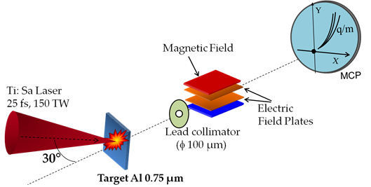
Fig. 1: The schematic experimental diagram is shown in (a). The Thomson Parabola Ion Spectrograph (TPIS) is used as primary diagnostics for ion emission studies.
Typical ion signal recorded using TPIS is shown in Fig 2 (a). Derived ion energy spectra is shown in Fig. 2(b). To record the proton beam profile, a stack of RCF was placed behind the target. Fig. 2 (c) shows the proton beam imprint on different layer of RCF. The respective energy cut-off for each layer is also shown below. 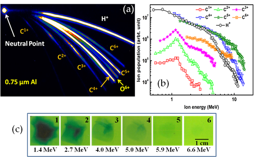
Fig. 2: (a) Typical TPIS trace observed in experiment. (b) Retrieved ion energy spectra and (c) proton beam profile recorded on RCF stack.
- Study of nuclear reactions using laser accelerated ion beam
Short lived radioisotope 11C and 18F have important applications in Positron Emission Tomography (PET). It is a non invasive diagnostic imaging technique which measures the metabolic activity of cells in human body. Short lived positron emitting isotope is injected into the body which further gets concentrated in tissue of interest. PET then construct the three dimensional image of the body by tracing the radioisotope that was injected in form of medicine. The PET isotope have half life few tense of minutes, therefore they need to be produced near the place of their use. Conventionally, ion beam from cyclotrons or linear accelerators are used to produce the PET isotope. This requires considerably large infrastructure in terms of accelerator cost and massive shielding. In the present work we tried to study the productions of 11C isotope by laser accelerated proton and deuteron beam using 11B(p,n)11C and 10B(d,n)11C reactions. We found that the laser driven ion beam can produce substantial activity and be a viable alternative in future with further advances in laser technology.
Experiment has been performed using 25 fs, 150 TW Ti:Sa laser system. The laser pulse is focussed onto the thin foils using a 30° off axis parabolic mirror on to the target resulting an intensity of ~ 1×10²⁰ W/cm2. Foils of different materials and thickness like 1.5 µm Ni, 1 µm Cu, 0.75 µm Al, CD2 coated Ni foil, 8 µm Plain CH foil and 8 µm CD2 foil have been used as target. Protons (H+) are accelerated from the rear surface of thin foil. In case plain CD2 and CD2 coated Ni foil high flux of deuterium ions was also present along with other ions. Protons up to 8 MeV and deuterium ions up to 5 MeV could be accelerated. A 2 mm thick 99.9 % pure natural Boron (in natural Boron ; 80% 11B and 20% 10B) palette is then kept in the ion beam path for reactions to occur. The experimental schematic is shown in Fig. 3(a).
After irradiation with proton/deuteron beam, the generated 11C activity was recorded using High Purity Germanium detector (HPGe). The 11C decays into 11C→11B + e+ + νe. The positron annihilates with background electron and gives two gamma photons of 511 keV. The gamma spectrum recorded using HPGe detector shows a strong peak at 511 keV(Fig 3b), indicating the substantial generated 11C activity in the boron sample. The measured half-life of the11C isotope was found to be 20 min which is very close to the actual value 20.3 min (inset of Fig 3b). In optimized condition and at ~ 100 TW laser power the produced activity per shot was 3.5 ×10³ Bq (for Cu 1 µm target and using 11B(p,n)11C). We found that the use of deuteron beam can further enhance the overall activity of 11C isotope because of the low energy threshold of the reaction 10B(d,n)11C.
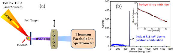
Fig. 3: (a) Experimental set up (b) Gamma spectrum and half life measurement using HPGe detector.
Necessary dose for PET applications is ~ 200 MBq, which could be possible to achieve using a laser operating at 100 Hz, for 600 seconds under present experimental condition, for which cumulative activity can reach up to 210 MBq. The results offer a promising source of PET isotope generation based on ultrashort pulse laser driven ion beam which looks to be feasible with further advancement in laser technology.
- Ion beam handling and transport
Presently laser ion acceleration has few draw backs. The accelerated ion beam exhibit broad energy distribution and ions are emitted in a cone with typical opening angle of ~10°-15°. To overcome this problem and enable the beam utilization at far of distances from the source, researchers are adopting the hybrid approach i.e., after initial acceleration stage use of conventional ion optics for beam transport and manipulation. Currently several projects on beam line development for laser accelerated ion beam source are being pursued across the world. Recently, at Laser Plasma Division a high field pulsed solenoid base magnetic lens is developed to focus /collimate and transport the beam for radiography application involving physical structures and living cells. Here a brief account of the experimental detail and result is presented.
The experimental scheme is shown in Fig. 4 Laser pulses from 150TW, Ti:sapphire laser system were focused on to the thin foil target using an Off axis parabolic mirror to a spot size (FWHM) of ~ 6 µm resulting in peak laser intensity of ~ 1020 W/cm2. 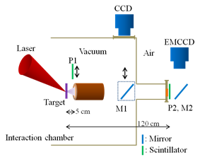
Fig. 4: Experimental scheme showing laser ion acceleration from thin foil target and subsequent capture and transport using solenoid
Ions are emitted from foil rear surface along normal direction in a certain cone angle which reduces for high energy ions. The solenoid having clear inner diameter of 32 mm effective length 16 cm. It was derived using in house developed pulsed power supply based on capacitor discharge scheme. The solenoid current vs field characteristic was studied in detail prior to its use in the experiment.
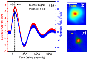
Fig. 5: (a) Current vs magnetic field characteristic of the pulse solenoid. (b) Proton beam profile before the solenoid. (c) Focused beam profile in air at 1.2-meter distance from the source.
The deriving current profile and the resultant magnetic field is shown in Fig. 5 (a). At 7.5 kA current around 5 T magnetic field was produced inside the solenoid. The solenoid operation was synchronized with laser operation such that the accelerated proton beam observes the peak field as it passes through solenoid (indicated by vertical dotted lines). The proton beam profile was monitored online at different positions using 250 µm thick plastic Scintillator (EJ 228) and CCD camera combination. The Scintillator was covered with appropriate Al foils having different energy cut-off. Fig. 5 (b) shows proton beam profile at 5cm distance from the source i.e. just at the input of the solenoid. The diverging nature of the proton beam is apparent. As the ion beam has broad energy distribution, therefore different energy protons get focused at different positions. It was observed that at 5 T field, the 4 MeV protons are focused at 1.2 m distance from the source. Fig. 5 (c) shows focused beam profile of 4 MeV protons at focused position (P2). The beam is taken out from vacuum chamber through 50 µm thick kapton window. We had also estimated the transfer efficiency of the solenoid by estimating the input and output proton beam flux using CR-39 detector. It was found that about 108 protons of 4 MeV energy with around 30% efficiency could be transported to position P2 in a single laser shot.
- Radiography of static microstructures and highly transient fields inside plasma
Laser accelerated proton beam have been used for radiography of physical microstructures and expanding plasma. Main compressed pulse of 25 fs pulse duration 1020 W/cm2 intensity was used to generate proton beam up to ~ 4MeV energy from thin foil targets. As shown in Fig 6 (a), the proton beam was directly used for radiography by placing the object (microstructure/plasma) at few mm distance from the source. The imaging magnification was 10-15x. For plasma radiography a separate pre-pulse of 500 ps pulse duration was used to generate the pre-plasma on a knife edge target. The plasma evolution was probed at different times by adjusting the time delay between the main pulse (proton producing) and pre-pulse. The radiographic images were recorded on Radiochromic Films (RCF) and Fast scintillator and CCD camera combination. A typical radiography image of a TEM grid (20-micron bar width and 70 microns spacing at 10x magnification) is shown in Fig 6 (b). The radiography of expanding plasma at few hundred ps time steps recorded on RCF is also shown in Fig 6 (c). The protons being charge particles get deflected by the azimuthal magnetic field inside the plasma as clearly seen in the radiographic images.
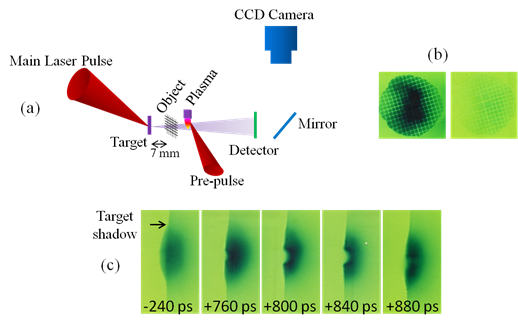
Fig. 6: (a) Schematic of radiography set up. Radiograph of a (b) TEM mesh and (c) expanding plasma produced by sub ns laser pulse.
Accelerated ion beam has been envisaged as an important source in radiation oncology because of its ability to impart localized energy deposition without collateral damage unlike photon and electron beams. The protons and lighter ions e.g. carbon, deliver most of their energy at the end of their path, at the so-called Bragg peak. This property makes protons and lighter ions very suitable for highly localized energy deposition. The conventional hadron therapy facility based on rf accelerator based source is large and very expensive. Major part of it goes in transport of the particle beam to the patient (40 – 50 %). The Laser based proton / ion sources offers a promising alternative. The beam transport cost can be drastically reduced as the ion source can be produced very close to the patient by laser beam. In our recent study using 0.4 μm thick Al foil we have now able to record protons up to 11 MeV and C4+ up to 14 MeV energies. It is expected that with improvised target geometries and proper conditioning of the laser pulse, a considerable increase in proton and heavy ion energy and flux will be accomplished which will be quite useful to study the hadron therapy.
For more detail please see the publications appearing on the website.
|