In recent years, Raman spectroscopy has been suggested and validated as a potential tool for non-invasive and near real-time diagnosis of various tissue abnormalities. We have an ongoing activity on Raman spectroscopy for various biomedical applications.
(1) In-vivo Raman Spectroscopy for Detection of Oral Neoplasia
We carried out detailed studies to evaluate the applicability of in-vivo Raman spectroscopy for differential diagnosis of malignant and potentially malignant lesions of human oral cavity in a clinical setting. The in-vivo studies were conducted at Tata Memorial Hospital (TMH), Mumbai using the portable Raman spectroscopic system developed at LBAD.
The Raman spectra, measured from multiple sites of normal oral mucosa and of lesions belonging to either of the three histopathological categories, viz. oral squamous cell carcinoma (OSCC), oral submucous fibrosis (OSMF) and leukoplakia (OLK), were subjected to a probability based multivariate statistical algorithm capable of direct multi-class classification. In comparison to histology, the diagnostic algorithm was found to provide an accuracy of 85%, 89%, 85% and 82% in classifying the oral tissue spectra into the four different pathology classes: normal squamous tissue, OSCC, OSMF, and OLK respectively, along with an overall accuracy of 86% based on leave-one-individual-out cross validation. When employed for binary classification, the algorithm resulted in a sensitivity and specificity of ~94% in delineating normal from the rest of the abnormal spectra of OSCC, OSMF and OLK tissue sites pooled together.
(a) Portable Raman Spectroscopy system developed at LBAD, (b) Mean, normalized Raman spectra of oral tissues belonging to different pathologies. Ref: Journal of Biophotonics, 7(9), 690-702, 2014. https://doi.org/10.1002/jbio.201300030
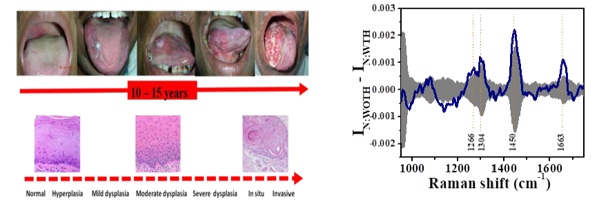
The in-vivo Raman spectra recorded from healthy volunteers were also analyzed to explore whether the sets of spectra were different amongst those with tobacco habits and those without any tobacco habits and the effect of these changes on differential diagnosis of oral lesions. It was found that the Raman spectra of healthy volunteers with tobacco consumption habits could be separated from the spectra of those without any habit of tobacco consumption with an accuracy of over 95%. Further, it was found that exclusion of the spectral data of the oral cavity of the healthy volunteers with tobacco habits from the reference normal database considerably improved the overall classification accuracy (92% as against 86%) of the algorithm in separating the different oral lesions from the normal oral tissues.
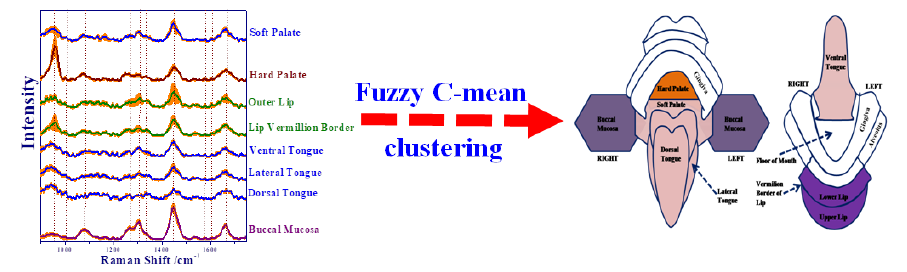
Clinical studies were carried out to characterize the variability of Raman spectra measured in-vivo from the different anatomical locations of the oral cavity of healthy volunteers and investigate its effect on the outcome of statistical discrimination of malignant and potentially malignant oral lesions from the healthy oral mucosa. An unsupervised cluster analysis using Fuzzy c-means clustering algorithm was conducted for quantifying the underlying structure of the normal oral tissue spectra. The algorithm could segment the normal oral tissue sites, based on similarity of spectral patterns, into four major anatomical clusters (AC): (1) outer lip, and lip vermillion border into AC-I with an accuracy of 80%; (2) buccal mucosa into AC-II with an accuracy of 72%; (3) hard palate into AC-III with an accuracy of 92%; (4) dorsal, lateral and ventral tongue and soft palate into AC-IV with an accuracy of 76%. A probabilistic multi-class diagnostic algorithm, developed for supervised classification, was used to classify the whole set of measured tissue Raman spectra into three categories: normal, potentially malignant (OSMF and OLK pooled together) and malignant (OSCC). The results showed that the diagnostic algorithm, when applied on the pooled set of spectra from all the anatomical clusters, correctly discriminated normal, malignant and potentially malignant tissue sites with 86%, 88%, and 86% accuracy respectively, which amounted to an overall accuracy of 87%. However, when the anatomy-matched data sets were considered, the overall classification accuracy was found to improve to 95% with the algorithm correctly discriminating the corresponding tissue sites with 94%, 99%, and 91% accuracy, respectively.
Clustering of Raman spectra based on the anatomical locations of the target tissue sites of the oral cavity. Ref: Biomedical Spectroscopy and Imaging, 2(13), 199-217, 2013.
Progression of oral cavity cancer with tobacco consumption habits and the mean difference spectra showing statistical differences between oral tissue Raman spectra of healthy volunteers without any tobacco consumption habit (N:WOTH) and with tobacco consumption habit (N:WTH). Ref: Journal of Analytical Oncology: 5(3), 110-123, 2016. http://dx.doi.org/10.6000/1927-7229.2016.05.03.4
(2) Development of range-independent background subtraction algorithm (RIA)
Central to clinical Raman spectroscopy for tissue diagnostic applications is an appropriate algorithm that can faithfully retrieve weak tissue Raman signals from the intense background of the measured raw Raman spectra.
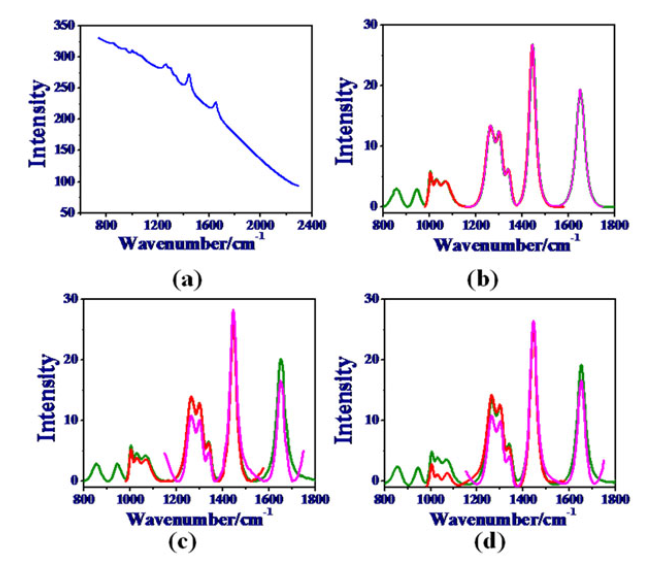
The most widely used method for background subtraction is iterative polynomial fitting to mimic the contour of the background. However, in this method, the lineshape and intensity of the extracted Raman spectra are found to be dependent on the range of the wavenumbers selected for the fitting, thereby leading to significantly different Raman spectra for different start and stop wavenumber selections. We have developed a novel background subtraction algorithm based on iterative smoothening of the raw tissue spectra which overcomes this drawback and gives range independent Raman spectra. Given a raw Raman spectrum and the choice of the start and the stop wavenumbers, the algorithm first truncates the spectrum to include the raw data within this wavenumber range, linearly extrapolates the truncated raw spectrum beyond the points of truncation on the two sides by using coefficients of linear least-square fit, adds two Gaussian peaks of appropriate height and width on the extrapolated linear wings on either side and then iteratively smoothens the data with all these add-ons such that the smaller of the ordinate values of the smoothed and the starting raw data serve as the input to each successive round of iterative smoothing until the added Gaussian peaks are fully recovered. The algorithm was compared with the modified polynomial-based algorithms using mathematically simulated Raman spectrum as well as experimentally measured Raman spectra from various biological samples and was found to yield consistently range-independent and artifact-free Raman signal with zero baseline.
(a) Raw Raman spectrum and the Raman signals recovered from it using (b) the RIA, (c) the ModPoly, and (d) the I-ModPoly for three different spectral ranges: (i) range-1 corresponding to 800-1800 cm-1, (magenta) (ii) range-2 corresponding to 980-1580 cm-1 (red), and (iii) range-3 corresponding to 1150-1750 cm-1 (green). Ref: Journal of Raman spectroscopy, 43(12), 1884–1894, (2012). https://doi.org/10.1002/jrs.4127
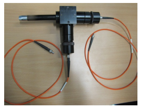
(3) Development of Raman probe for in-situ measurement of artefact-free Raman spectra from biological tissues
A Raman probe is an important component of the clinical Raman system intended for non-invasive in-vivo tissue analysis. However, the commercially available Raman probes are not optimally suited for studying the Raman characteristics of tissues for in-vivo diagnosis of a disease. The Raman probe developed at RRCAT is meant for in situ measurement of good quality Raman spectra from low Raman-active materials like biological tissues.
Unlike the commercially available Raman probes which are found to introduce various artefacts that interfere with the Raman signatures appearing in the fingerprint region of the tissue Raman spectra and lead to confusion in its interpretation, the developed probe is based on a novel design by which it helps avoid these artefacts. Further, the design also optimizes collection efficiency enabling in situ measurement of good-quality, artefact-free Raman spectra from biological tissues within a few seconds.
The technology of the developed Raman probe has been transferred to M/s Applied Optical Technologies Pvt. Ltd., Ambernath, Thane. It is expected to further the use of Raman spectroscopy in the field of biomedical applications where in situ measurement of Raman spectra is often desired but not possible because of unavailability of a suitable Raman probe.
Raman probe developed at LBAD
(4) Depth-sensitive Raman spectroscopy
In the typical configuration of Raman spectroscopy, a given tissue sample is illuminated by a laser of appropriate wavelength and the Raman signal back-scattered from the tissue is detected by a Raman spectrometer for further analysis. Though this kind of conventional measurement has been shown to be useful in certain situations, its major shortcoming is that it obtains information only from the surface of a tissue. However, the majority of biological tissues are known to have sub-surface layers, with different layers having different biochemical and morphological make-up. Further, the morphological and biochemical changes, that these sub-surface layers undergo as tissue transforms from normal to diseased, are also believed to be quite different from that of the surface. Consequently, the resulting changes in the optical properties that get manifested in the Raman signatures of the surface and the sub-surface tissue layers are not expected to be the same. Since in the conventional approach, the measured Raman signal at a given point on the surface of an interrogated tissue is volume integrated (over the sub-surface depths), it does not contain the desired information of the sub-surface tissue layers having different Raman characteristics. Obtaining depth-wise Raman signal is important because it may allow for a more detailed analysis of the biochemical (and morphological) state of a given tissue thereby leading to an improved feedback on tissue state.
Our ongoing research on depth-sensitive Raman spectroscopy for the analysis of layered turbid media has resulted in the development of various novel approaches of depth-sensitive Raman measurements from tissues as listed below.
Off-confocal Raman spectroscopy (OCRS): The method uses the experimental configuration of a confocal Raman, but employs off-confocal Raman detection for sub-surface interrogation of layered tissues.
The technique allows subsurface interrogation by moving the tip of the Raman detection fiber (acting as the pinhole aperture) from the focus of the Raman collection objective either by taking the point of detection away from the objective (positive confocal offset) or bringing it closer to the objective (negative confocal offset). The off-confocal separation of the detection point leads to micrometer-scale spatial offset between the collection and the fixed-point illumination on the surface of the target sample, thereby enabling to probe subsurface layers inside the sample at depths beyond the reach of the conventional confocal Raman. The two important advantages of this approach are that it does not require any adjustment in the sample arm of the system and the illumination light remains fixed on the sample surface and thereby providing the effective illumination. A depth-sensitive Raman probe based on this principle has also been developed which is undergoing validation.
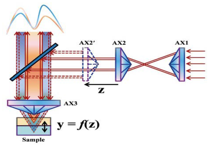
Cone-Shell Raman Spectroscopy (CSRS): Though the off-confocal Raman spectroscopy approach was found to solve the practical limitations of confocal Raman spectroscopy in probing sub-surface layers, yet the depths beyond ~800 μm could not be probed using OCRS. For measuring Raman signals from a depth beyond this limit, another new depth-sensitive Raman spectroscopic technique using the configuration of cone-shell Raman excitation and detection was developed.
The technique was named cone-shell Raman spectroscopy (CSRS). The CSRS system uses a 785 nm diode laser and three identical axicons for Raman excitation of the target sample in the form of a hollow conic section. The Raman light, backscattered from the sample and passed through the same conic section, is collected for detection. An important attraction of the system is that the probing depth can be varied by simply varying the separation between axicons in the excitation arm. No adjustment is required in the sample arm, which is a significant advantage, similar to the case of OCRS, for non-contact, depth-sensitive measurement. Using this approach, Raman signal from a depth of up to few mm (~2.5 mm) beneath the surface can be measured.
Schematic of the experimental setup for cone–shell Raman spectroscopy (CSRS). Ref: Journal of Biophotonics, 8(1-12), 889-896, 2015.
https://doi.org/10.1002/jbio.201400125
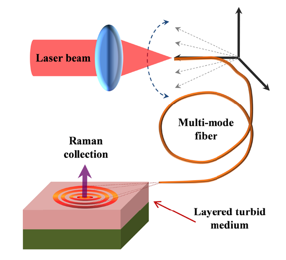
Axicon lens free scheme for implementing inverse SORS: In this scheme, unlike the conventional inverse SORS based setup where an axicon lens is used for obtaining a ring shaped illumination beam, such light beam is generated using a multi-mode fiber (lying in the source arm of the experimental set-up) kept at non-zero angle with respect to the axis of the lens used for focusing the laser light onto its tip.
On varying this angle, the Raman illumination beam is incident onto the sample surface in the form of concentric illumination rings of varying radii. The back-scattered Raman light exiting the sample surface at the centre of the concentric rings is collected for the detection. This approach not only probes higher depths as compared to CSRS but would allow one developing a compact and pencil-sized SORS probe, an urgent and current need in routine clinical pathology setting.
(5) Detection of analytes in body fluids for biomedical diagnosis
Accurate and sensitive detection of disease-specific analytes present in body-fluids plays a crucial role in disease diagnosis and prognosis. Recently, Raman spectroscopy has garnered a great deal of interest in the analyses of body-fluids.
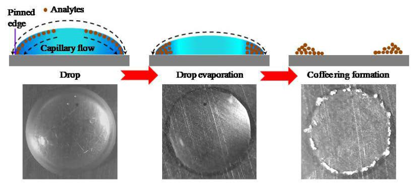
Its attractiveness comes from its ability to probe molecular specific information of each of the constituent analytes present in a body fluid in greater detail than other spectroscopic techniques like absorption or fluorescence. However, the conventional Raman spectroscopy does not have enough sensitivity in detecting the typical concentrations at which most of the analytes are present in body-fluids. Efforts are, therefore, ongoing to overcome this limitation and enhance the Raman signal so as to be able to detect the bio-analytes at their physiological concentrations. At LBAD we are working on different techniques for enhancing the Raman signal and also evaluating the basic challenges and limitations of these techniques and how they apply to the detection of bio-analytes. For example we have evaluated the applicability of drop-coating deposition Raman spectroscopy (DCDRS), a relatively recent variant of Raman spectroscopy, for quantitative determination of creatinine present in urine. The technique relies on the formation of a “coffee ring” pattern by drying up of a microliter volume drop of a liquid containing an aqueous solution of the analyte and then, detecting the Raman signal from the dried-up pattern of this drop. The results of our study show that this technique can detect creatinine present in the artificial urine samples with an accuracy of over 94% in the physiological concentration range.
Mechanism of Drop Coating Deposition. During the drying of a sessile drop, capillary flow results in deposition of solutes at the edge to form a coffee ring. Top panel is schematic representation of the process. Bottom panel shows bright-field images of the drop of an aqueous solution of an analyte at different time points during drying.
(6) Raman optical tweezers
Microscopy based imaging and spectroscopy at cellular level is an important tool for understanding the basis of biomedical diagnosis and therapy. AT LBAD, we have developed (holographic) optical tweezers and Raman optical tweezers for that purpose.
A variety of experiments were carried out to demonstrate the potential of optical tweezers for biomedical applications. For example, it was shown that combining image analysis methods with holographic tweezers can successfully sort between red and white blood cells inside human blood samples. The other studies carried out with the holographic tweezers include use of three dimensional interference patterns among phase-engineered beams to manipulate objects over a long distance (~ 10 mm), an essential requirement for successful integration of optical traps with microfluidic devices.
Raman optical tweezers involves recording of Raman spectra from optically trapped biological cells. The technique being non-contact in nature causes minimal perturbation to cells under investigation, and offers improved signal to noise ratio for the spectra. In one of the studies, we investigated the photo induced oxygenation state changes of human red blood cells (RBCs) as may happen under optical trap when the laser power is not chosen judiciously. In a more recent study, we investigated the effect of hypoxic exposure on RBCs as may occur at high altitudes and conditions like cardiac failure, obstructive pulmonary disease etc. The results of this study show that when RBCs are kept in an oxygen deprived condition for increasing durations, a reducing hemoglobin (Hb)-oxygen affinity results for the cells. In another study, we investigated the changes in RBCs when undergoing common shape transformations (like transformations of normal discocytes to echinocytes and stomatocytes) seen under diseased states or during interaction with various drugs. It was found that compared to discocytes and stomatocytes the echinocytes have an increased level of Hb-oxygen affinity, detrimental for effective release of oxygen at tissue sites. Also the echinocytes showed sign of Hb damage when deoxygenated. We also performed studies on assessing the effects of low level microwave radiations, widely used in mobile phones, on RBCs. Since, by now, the possible role of mobile radiation in the development of malignant type of brain cancer (glioma) is quite accepted, we carried out studies on RBCs when exposed to varying duration of exposure from 900 MHz and 1800 MHz GSM mobile handset. Noticeable changes in the Raman spectra after exposure could be seen for cells under radiation for duration of 30 minutes or more, suggesting altered oxygen carrying capacity for the RBCs. Further, since it is known that in combination with spectroscopy and suitable chemometric methods Raman optical tweezers has the potential to be successfully applied to distinguish between malignant and healthy cells, we have initiated studies on known cancer cell lines to gain valuable understanding of the disease mechanisms at single cell level that might lead to future development of clinically useful diagnosis methods.
Schematic of Raman optical tweezers
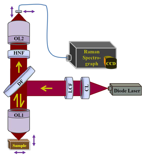
(7) Combined Raman spectroscopy and optical coherence tomography for tissue analysis
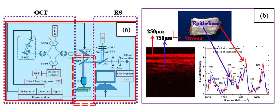
At LBAD, we have been actively pursuing the activity on depth-sensitive Raman spectroscopy and imaging for biomedical applications. The objective is to innovate on multimodal optical techniques to aid in the non-destructive and rapid characterization of the molecular, biochemical and structural properties of human tissues. Much of our currents efforts have been directed towards this objective. For example, recently we combined depth-sensitive Raman spectroscopy (RS) and optical coherence tomography (OCT) for revealing both biochemical and morphological structure of layered tissues. For achieving the depth-sensitivity in RS, we adopted a confocal configuration with a low NA (=0.4) objective lens. The objective lens was chosen in such a way so that both RS and OCT might be able to provide almost equal probing depths (up to -1 mm) for a two layered tissue comprising ~250 μm thick epithelial over a relatively thick stromal layer. The use of a low NA objective lens, though, degraded the depth resolution of the RS to a value of ~210 μm, it was enough to separate the Raman signatures of the individual tissue layers. In a similar manner, we also combined the OCT with a depth-sensitive fluorescence set up developed using a low NA objective lens.
(a) The schematics of the combined Raman-OCT system developed at LBAD (b) the OCT image of a two layered biological tissue along with the Raman spectra of different tissue layers. Ref: Journal of Biophotonics, 7, 77-85, (2014).
https://doi.org/10.1002/jbio.201200208


 Progression of oral cavity cancer with tobacco consumption habits and the mean difference spectra showing statistical differences between oral tissue Raman spectra of healthy volunteers without any tobacco consumption habit (N:WOTH) and with tobacco consumption habit (N:WTH). Ref: Journal of Analytical Oncology: 5(3), 110-123, 2016.