X-ray fluorescence (XRF) spectroscopy is a powerful non-destructive method for micro and trace elemental characterization of materials. The technique is used in a range of domains, including geology, archaeology, biomedical science, and material science, etc. The BL-16 beamline allows a variety of re-configurable operational modes (normal XRF, TXRF, and micro-XRF modes) with minimal set-up time. It provides an attractive platform for researchers to conduct a wide range of research activities, particularly in the fields of archaeology, earth science, and environmental applications, etc.
Beamline parameters & Optical layout
Parameters
Values
Source
Bending magnet
Energy Range
5-20 keV
Beam acceptance
1 mrad (h) × 0.2 mrad (v)
Energy resolution(ΔE/E)
~ 10-3-10-4
Beam spot size (at sample position)
~ 7.5 µm (h) × 4.3 µm (v) [Focused mode]
~ 22 mm (h) × 5 mm (v) [Unfocused mode]
Monochromator
Si (111) double-crystal monochromator
Photon Flux [at 10 keV X-rays/ 100 mA ring current]
~ 2 × 10⁷ photons/sec [Focused mode]
~ 1 × 10⁸ photons/sec/mm2 [Unfocused mode]
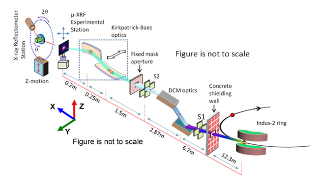
BL-16 Beamline optical layout
The BL-16 beamline uses a fixed-exit double crystal monochromator (DCM) (FMB, Berlin, Germany) with a pair of symmetric and asymmetric Si (111) crystals (mounted side-by-side).
The beamline utilises various types of slit systems (water cooled and un-cooled) for controlling the final beam size available at the experimental station, as per the user requirements.
The beamline also utilises a Kirkpatrick–Baez (KB) optics (Xradia, USA), comprising of two elliptical bendable mirrors for focusing of the synchrotron X-ray beam.
The beamline offers minimum micro focus beam dimensions ~ 5 μm (V) × 8 μm (H) for microfluorescence mapping applications.
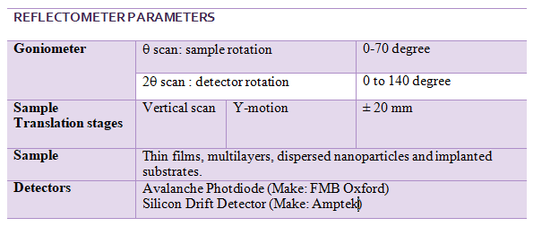
The BL-16 beamline also includes a third experimental station for analysis of thin layered materials using X-ray reflectivity (XRR) and grazing incidence X-ray fluorescence (GIXRF) measurements.
Experimental station
The BL-16 beamline allows various re-configurable experimental stations (for normal XRF, TXRF, micro-XRF, XRR and GIXRF measurements) with a minimal set-up time, which allows a user to perform a wide range of experiments.
An inside view of the experimental hutch of the BL-16 Beamline
Experimental facilities
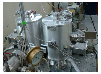
1) XRF-TXRF chambers
This setup is used for the XRF and TXRF analysis of different types of samples (i.e. bulk, liquid and powder forms).
XRF-TXRF chambers
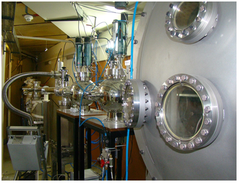
2) Two-Circle Goniometer
This setup is used for simultaneous X-Ray Reflectivity (XRR) and Grazing Incidence X-ray Fuorescence (GIXRF) of thinflims, multilayers and nano structured materials.
A photograph of the BL-16 Goniometer setup
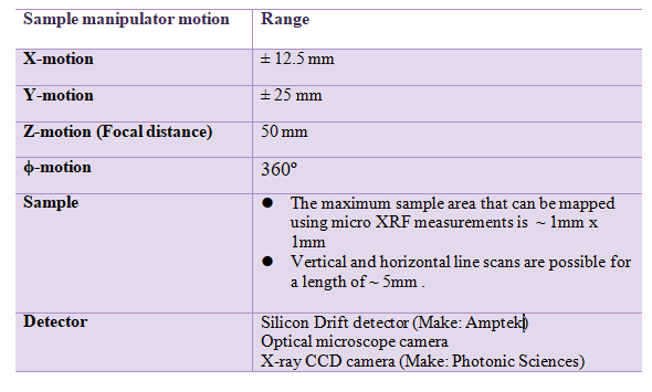
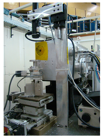
3. MicroXRF
This setup is used for the elemental mapping of heterogeneous multi-element samples using micro focused X-ray beam. A five-axis sample manipulator stage is used for mounting of the sample.
A photograph of the BL-16 MicroXRF sample manipulator system
Application Areas
The different experimental stations of the beamline provide an attractive platform for conducting a wide range of research activities, particularly in the fields of archaeology, earth science and environmental applications, and materials science. Some of the research fields are given below.
Materials and Engineering
Thin films, multilayers and nanoparticles
Elemental analysis of Medicines
Environmental monitoring and toxicity
Life science
Food, Agricultural & Geological materials
Forensic applications
1.
“Structure, stress, and optical property correlations in nano-structured Mo/Si and W/B4C multilayer mirrors for hard x-ray applications”
Faiyaz Mollick, M. Nayak, Ajay Kumar Kashyap, Jitendra Kumar, Arindam Majhi, Nageswararao Pothana, Parasmani Rajput, M. K. Tiwari, Sanjay Kumar Rai, Manvendra Narayan Singh, Archna Sagdeo
Journal of Applied Physics, 138(17) (2025).DOI: 10.1063/5.0291236.
2.
“Identification of transition metals and their alloys using a synchrotron X-ray source and a MOVPE grown GaAs p-i-n detector”
Geetanjali Vashisht, Payal Taya,Ravi Kumar, Ayushi Trivedi, M. K. Tiwari, Tarun Sharma, Vijay Kumar Dixit
Nuclear Instruments and Methods in Physics Research Section A, 1084:171184 (2025).DOI: 10.1016/j.nima.2025.171184.
3.
“Analysis of TiO2 and Ti Nanolayers Irradiated With Highly Charged Xe q+ Ions Using Synchrotron Radiation Based GIXRF Method”
Regina Stachura, Dariusz Banaś, Aldona Kubala-Kukuś, Paweł Jagodziński, Paweł Jagodziński, M. Pajek, Karol Szary, Ilona Stabrawa, Weronika Biela, Giuliana Aquilanti, Iva Božičević Mihalić, Mohammad Akhlak Alam, M. K. Tiwari, M. Teodorczyk
X-Ray Spectrometry, 55 (1), (2025).DOI: 10.1002/xrs.70029.
4.
“Enhancing the photocatalytic water electrolysis performance of Zn2 SnO4 nanostructures via post-synthesis nitrogen doping”
Lokanath Mohapatra, Akshay Kumar Sonwane, Sonali Samal, Tushar Chauhan, Parveen Garg, Uday Deshpande, M. K. Tiwari, Ajay Kumar Kushwaha
Nanoscale, 17 (32), (2025).DOI: 10.1039/D5NR01576F.
5.
“Investigations on the effect of arsenic and phosphorus atomic exchange on the origin of crystal potential fluctuations in InAsP/InP epilayers”
Geetanjali Vashisht, R. Roychowdhury, P. Rajput, R. Kumar, A. Trivedi, M. Balal, A. Bose, M. K. Tiwari, S. R. Barman, T. K. Sharma and V. K. Dixit
Applied Surface Science, Vol 682, 161627 (2025).
1.
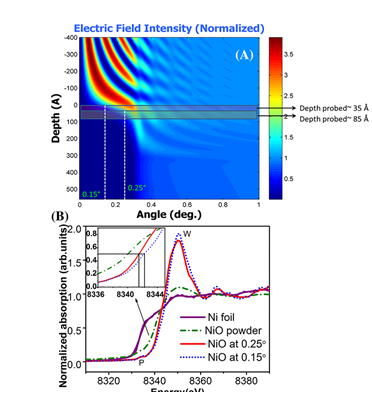
“X-ray standing wave assisted XANES for depth dependent chemical state analysis of Cr in Cr2O3/Cr bilayer structure” Ayushi Trivedi, Akhlak Alam, Ajay Khooha, Rajnish Dhawan, Rajendra Sharma, Shilpa Tripathi, M. K. Tiwari,
, Surface and Interface Analysis, 2024; 56(11): 760-769.doi:10.1002/sia.7342.
1.
Improvement of limit of detection sensitivities in the parts per billion range using conventional geometry synchrotron radiation excited EDXRF measurements Md. Akhlak Alam, M.K. Tiwari, Ayushi Trivedi, Ajay Khooha and A. K. Singh
J. Anal. At. Spectrom., Vol. 37, pp 575–583 (2022).
DOI: 10.1039/d2ja00016d
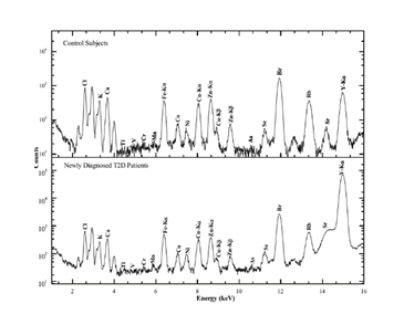
Serum Elemental Analysis of Type 2 Diabetes Patients Using SRXRF N. Srinivasa Rao, G. J. Naga Raju, M. K. Tiwari, B. G. Naidu, P. Sarita
Biological Trace Element Research, volume 200, pp 1485–1494 (2022).
https://doi.org/10.1007/s12011-021-02762-7.
1.
Near edge absorption studies of pure and impure NbSe2; theory and experiment Soumyadeep Ghosh, Rukshana Pervin, Haranath Ghosh, M. K. Tiwari, Parasharam Shirage
Journal of Materials Science, Vol. 56, 17062–17079 (2021).https://doi.org/10.1007/s10853-021-06375-2
2.
Assessment of the Elemental Profile of Leafy Vegetables by Synchrotron -Radiation Induced Energy Dispersive X-Ray Fluorescence Spectroscopy AS Bharti, S Sharma, A K Singh, M K Tiwari, K N Uttami
Journal of Applied Spectroscopy Vol. 88, 653-661, (2021).https://doi.org/10.1007/s10812-021-01221-4
3.
Synchrotron Based TXRF for Assessment of Treated Waste water VK Garg, AL Srivastav, MK Tiwari, A Sharma, VS Kanwar
Nature Environment and Pollution Technology , Vol 20 (2), 743-746 (2021)https://doi.org/10.46488/NEPT.2021.v20i02.034
1.
Elemental analysis of Arsenicum album 30 KA K Dwivedi and M K Tiwari
International Journal of Homoeopathic Sciences, Vol. 4(4), pp 135-137 (2020).
2.
Solar X-Ray Monitor on Board the Chandrayaan-2 Orbiter: In-Flight Performance and Science Prospects N. P. S. Mithun et. al.
Solar Physics, Vol. 295 , Article number: 139 (2020) .https://doi.org/10.1007/s11207-020-01712-1
3.
Measurement of L subshell fluorescence yield ratios of some high Z elements by selective excitation method G. B. Hiremath, A. S. Bennal, M. M. Hosamani, N. M. Badiger, A. Trivedi, M. K. Tiwari
X-Ray Spectrometry , Vol 50, Issue1, pp 37-44 (2021) https://doi.org/10.1007/s11207-020- 01712-1
Study of the solid-state effect on L3 subshell fluorescence yield for high Z targets using Indus-2 synchrotron radiation Gangadharayya B. Hiremath, A.S. Bennal, Santosh Mirji, M.M. Hosamani, N.M. Badiger, and M. K. Tiwari,
Canadian Journal of Physics, Vol. 98(5):470–473 (2020).https://doi.org/10.1139/cjp-2019-0252
1.
Prompt Screening of the Alterations in Biochemical and Mineral Profile of Wheat Plants Treated with Chromium Using Attenuated Total Reflectance Fourier Transform Infrared Spectroscopy and X-ray Fluorescence Excited by Synchrotron Radiation Sweta Sharma, A. K. Singh, M. K. Tiwari & K. N. Uttam.
Analytical Letters, Vol 53:3, 482-508 , (2019).https://doi.org/10.1080/00032719.2019.1656729
2.
Interface sharpening in miscible and isotopic multilayers: Role of short-circuit diffusion A. Tiwari, M. K. Tiwari, M. Gupta, H.-C. Wille,and A. Gupta,
Phys. Rev. B , Vol. 99, 205413 , (2019). https://doi.org/10.1103/PhysRevB.99.205413
3.
Morphological and elemental mapping of gallstones using synchrotron microtomography and synchrotron X‐ray fluorescence spectroscopy Mohana Bakthavatchalam, Jayanthi Venakataraman, Ramya J Ramana,Mayank Jain,
Balwant Singh, Arul K Thanigai, Vaithiswaran Velyoudam, Saravanan Manickam Neethirajan, Manoj K Tiwari, Ashish K Agarwal,and Narayana S Kalkura
Journal of gastroenterology and hepatology 3, pp 381-387 , (2019). https://doi.org/10.1002/jgh3.12171
4.
Multivariate analysis of trace elemental data obtained from blood serum of
breast cancer patients using SRXRF
B. Gowri Naidu, P. Sarita, G.J. Naga Raju, M.K. Tiwari
Results in Physics, Vol. 12, pp 673–680, (2019). https://doi.org/10.1016/j.rinp.2018.12.020
5.
Study the effect of crystal structure on radiative vacancy transfer
probabilities from L3to Mi,Ni and Oi subshells
Gangadharayya B. Hiremath, Santosh Mirji, M.M. Hosamani, N.M. Badiger,
M.K. Tiwari
Chemical Physics Letters, Vol. 715, pp 317-322, (2019). https://doi.org/10.1016/j.cplett.2018.11.058
1.
Effect of manganese stress on the mineral content of the leaves of wheat seedlings by use of X-ray fluorescence excited by synchrotron radiation Sweta Sharma, Abhi Sarika Bharti, M. K. Tiwari, and K. N. Uttam
Spectroscopy Letters Vol. 51, Issue 6, pp 302-310 , (2018). https://doi.org/10.1080/00387010.2018.1475399
2.
Fabrication and characterization of Er, Nd codoped Y2O3 transparent ceramic: A dual mode photoluminescence emitter Pratik Deshmukh, S. Satapathy, Anju Ahlawat, M.K. Tiwari, A.K. Karnal
Journal of Alloys and Compounds, Vol. 754, pp 32-38, (2018)https://doi.org/10.1016/j.jallcom.2018.04.297
3.
Total reflection X-ray Fluorescence determination of interfering elements
rubidium and uranium by profile fitting
Sangita Dhara, Ajay Khooha, Ajit Kumar Singh, M.K. Tiwari, N.L. Misra
Spectrochimica Acta Part B , Vol. 144, pp 87–91, (2018). https://doi.org/10.1016/j.sab.2018.03.011
4.
Recent trends in X-ray fluorescence spectrometry: precise investigation of
Nanomaterials
M K Tiwari
Spectroscopy Europe ,Vol. 30, No. 1, pp. 15-19 (2018) https://doi.org/10.1255/sew.2018.a1
5.
M sub-shell X-rayfluorescence cross-section measurements for six elements in the range Z=78–92 at tuned synchrotron photon energies5, 7 and 9 keV Himani Bansal, M.K. Tiwari, Raj Mittal
Journal of Quantitative Spectroscopy & Radiative Transfer,Vol. 204,232–241, (2018).https://doi.org/10.1016/j.jqsrt.2017.09.026
6.
An IAEA multi-technique X-ray spectrometry end station at Elettra Sincrotrone Trieste: bench-marking results and interdisciplinary applications Andreas Germanos Karydas, et al.
J. Synchrotron Rad., Vol. 25, pp 189–203, (2018). https://doi.org/10.1107/S1600577517016332
7.
Development of multilayer mirrors for space-based astronomical X-ray optics Singam S. Panini, M. Nayak, K. C. Shyama Narendranath, P. C. Pradhan, P. S. Athiray, P. Sreekumar, G. S. Lodha, M. K. Tiwari
Journal of Optics,Vol.47, Issue 1, pp 91–95, (2018).https://doi.org/10.1007/s12596-017-0444-8
1.
Elemental Investigation of the Leaf and Seed of Coriander Plant by Synchrotron Radiation X-ray Fluorescence Spectroscopy Abhi Sarika Bharti, Sweta Sharma, Nidhi Shukla, M. K. Tiwari, K. N. Uttam
National Academy Science Letters, Vol. 40, Issue 5, pp 373–377, (2017). https://doi.org/10.1007/s40009-017-0600-3
2.
Depth resolved chemical speciation of a W-B4C multilayer structure Gangadhar Das, A. G. Karydas, Haranath Ghosh, M. Czyzycki, A. Migliori, A. K. Sinha, M. K. Tiwari
Phys. Rev. B, Vol. 96, 155444, (2017).https://doi.org/10.1103/PhysRevB.96.155444
3.
Measurement of Coster-Kronig vacancy transfer factor of some lanthanides using monoenergetic X-ray photons
Krishnananda, Santosh Mirji, N.M. Badiger, M.K. Tiwari
Vacuum, Vol. 144, pp 160-163 ,(2017). https://doi.org/10.1016/j.vacuum.2017.07.031
4.
Determination of impurities in graphite using synchrotron radiation based
X-ray fluorescence spectrometry
M. Ghosha, K.K. Swain, P.S. Remya Devia, T.A. Chavan, A.K. Singh, M. K. Tiwari,
R. Verma
Applied Radiation and Isotopes, Vol. 128, pp 210–215 , (2017) https://doi.org/10.1016/j.apradiso.2017.07.025
5.
L X-ray intensity ratio measurements using selective L sub-shell photo-
ionisation on synchrotron
Himani Bansal, M. K. Tiwari, Raj Mittal
Radiation Physics and Chemistry, Vol. 139, pp 22–26 , (2017).https://doi.org/10.1016/j.radphyschem.2017.05.019
6.
Probing nanostructured materials using X-ray fluorescence analysis Gangadhar Das, Ajay Khooha, Ajit Kumar Singh, M. K. Tiwari
X-Ray Spectrom. 2017, Vol .46(5), pp 448–453, (2017) https://doi.org/10.1002/xrs.2777
7.
Measurement of L X-ray Fluorescence Cross-sections for 74W at Excitation Energies 12, 14, 15 and 16.5 keV with Synchrotron Radiation R. Kumar, A. Rani, R.M. Singh, M.K. Tiwari, A.K. Singh
Radiation Physics and Chemistry, Vol. 131, pp. 79-85 , (2017).https://doi.org/10.1016/j.radphyschem.2016.10.023
8.
Direct Determination of Oxidation States of Uranium in Mixed-Valent Uranium Oxides Using Total Reflection X-ray Fluorescence X-ray Absorption Near-Edge Spectroscopy Kaushik Sanyal, Ajay Khooha, Gangadhar Das, M. K. Tiwari, and N. L. Misra
Anal. Chem. , Vol 89 (1), pp. 871–876 , (2017).https://doi.org/10.1021/acs.analchem.6b03945
9.
L sub-shell fluorescence cross-section measurements for elements, Z = 62–67, at tuned photon energies Himani Bansal, M.K. Tiwari, Raj Mittal
Journal of Quantitative Spectroscopy and Radiative Transfer, Vol. 199, pp. 93–102 (2017).https://doi.org/10.1016/j.jqsrt.2017.05.007
Science Highlights
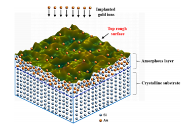
Depth profiling of energetic Au ions inside P-type Si 〈100〉 substrate
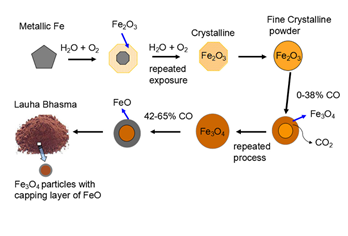
Structural investigation of Ayurveda Lauha (Iron) Bhasma
Journal of Ayurveda and Integrative Medicine, 14 100690 (2023)
Improvement of limit of detection sensitivities in the parts per billion range using conventional geometry synchrotron radiation excited EDXRF measurements