High brilliance, high coherence, and possibility of monochromatization at synchrotron sources has opened new horizons in X-ray imaging such as phase contrast imaging, diffraction enhanced imaging, real-time imaging and tomography of transient phenomena, and in-situ tomography under load conditions. X-ray phase contrast imaging has made it possible to distinguish features with very small density difference. The high intensity monochromatic beam also allows elemental mapping using K edge subtraction.
We have developed and installed an advanced imaging facility to carry out absorption and phase sensitive imaging and micro-tomography for material and bio-medical applications in research and industry. The beamline has white beam and monochromatic mode of operation. The maximum beam-dimension in the experimental station is 100 mm X 10 mm. Photon flux is ~1012 ph/s in monochromatic mode while it is 1016 ph/s in white-beam mode. Techniques such as radiography, propagation, analyzer and grating based phase contrast imaging, real-time imaging and tomography, in-situ imaging and tomography under load conditions etc. have been implemented. This facility is useful for advanced applications in bio-medical research and non-destructive characterization of advanced materials.
The Beamline outside view
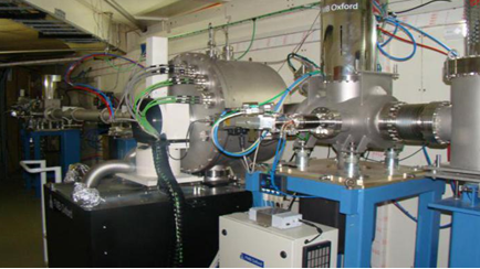
Inside view of optics hutch
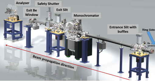
Optical layout & Beamline parameters
Parameters
Typical values
Source
Bending magnet, 1.5 T dipole
Operational modes
White and monochromatic
Monochromator
Si(111) Double crystal monochromator (DCM)
Energy range
8-35 keV
Energy resolution
(3.86x10⁻³ @ 12keV) in monochromatic mode
Beamline acceptance
5.5 mrad (H) × 0.5 mrad (V)
Photon flux
~ 108 ph/s/mm2 monochromatic energy;
~ 1016 ph/s (white beam)@ 2.5 GeV and 200 mA
Optics hutch comprises of components for beam shape and size selection, monochromatization, beam filtering, exposure and dose minimization etc. It consists of Water-cooled Be-window assembly, vertical and horizontal entrance slits, Si(111) Double Crystal Monochromator (DCM), vertical and horizontal exit slits, beam shutter and beam position monitor, water-cooled exit Be- window .
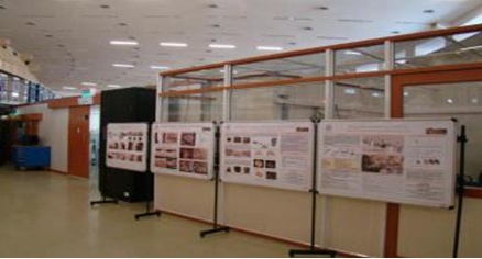
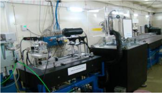
Experimental station
Photograph of the experimental station of imaging beamline
[1] Detectors-
X-ray imaging microscope: A lens coupled CCD detector with mirror optics and variety of combinations of scintillators and objectives for choosing required resolution (700 nm to 8 micron), field of view (1.5 mm to 16 mm)
High resolution X-ray CCD detector Fiber-Optic coupled, Gadox scintillator with 4007 × 2678, (18mm × 12mm), pixel size 4.5 micron.
A high speed imaging camera for Realtime imaging at microscopic resolutions with highest frame rate of 100 fps at full frame and up-to 1400 fps @ ROI of 2048 × 256
X-ray Flat panel detector for large area imaging :120mm field of view, 50 micron pixel size, 2400 × 2400 pixels
Silicon Drift detector for spectroscopic imaging and elemental mapping
[2] Motion stages -
High precision six axis sample manipulator stages consisting of (Y, Z, θ, ψ, φ)
High precision three-axis manipulator for detectors consisting X, Y, Z motions
[3] Other Instruments
Ion chamber for online beam current measurement and dose regulation
Fast shutter for controlled exposure time in bio-medical imaging.
Analyzer DCM, and diffraction gratings for differential phase and dark-field imaging.
500N and 3KN load cells for In-situ experiments under load conditions.
Complete experimental station on vibration isolated granite tables.
[4] Computational facilities
Computational facilities for image processing, micro-CT reconstruction, 3D visualization and quantitative analysis are available. Quantitative analysis for finding out structural features and density distribution in 2D and 3D is possible.
Experimental facilities
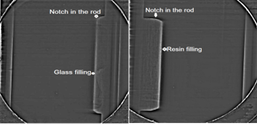
1) Absorption contrast imaging - 2D structure & Density mapping: Shape size and thickness - of the enclosed feature, particle, pore, voids, cracks, layers, coatings
Resin and glass filled notched features inside a 450 μm thick stainless steel simulant fuel rod
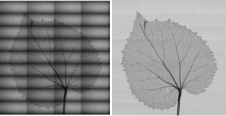
2) Large area imaging: High resolution 2D imaging of samples in size 150×90mm
Scanned ROIs of large samples leaf and stitched complete images
3) Phase contrast: 2D structure & Density mapping when density difference is small
Three type phase contrast techniques with different sensitivity and experimental/analytical complexity
In line phase contrast imaging
Phase contrast enhanced projection imaging
Diffraction enhanced imaging
Absorption, refraction and scattering contrast image.
Grating based phase contrast imaging
Quantitative phase contrast imaging with grating optics
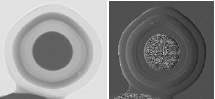
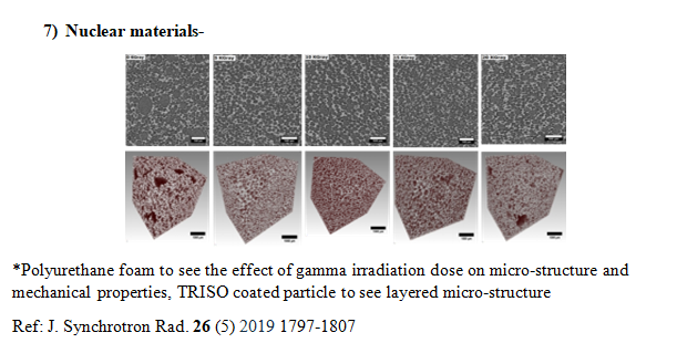
Visualization and measurement of layered micro-structure in TRISO fuel particle of CHTR
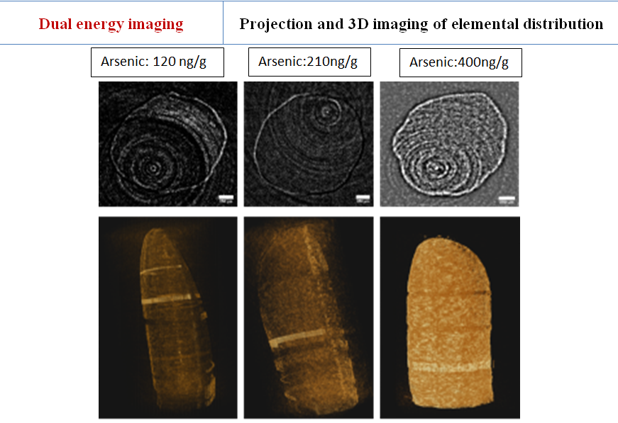
4. 2D & 3D elemental mapping
3D Mapping of Arsenic in Rice samples (concentration:120, 210 and 400 ng/g)
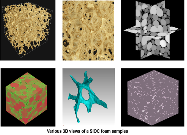
5. 3D structure & Density imaging
Various 3D view of SiOC foam sample generated using segmentation and colour coding
Localized micro-structural quantification of pores, cracks, shape, density etc
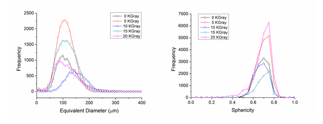
Quantitative measurement of porosity, and pore size in polyurethane foam irradiated with different gamma dose (J. Synchrotron Rad. 26 (5) 2019 1797-1807)
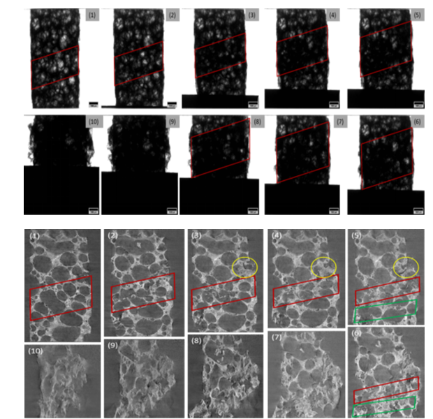
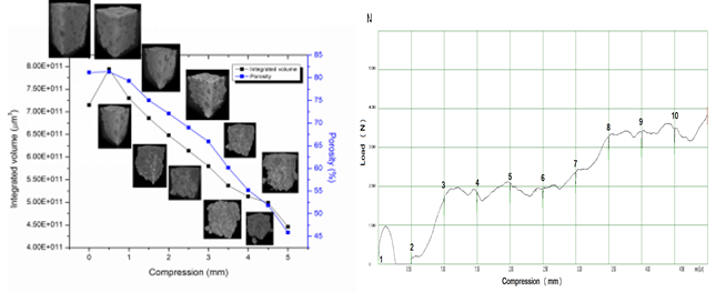
6. In-situ Imaging
In-situ deformation and stress band formation in Al foam under compression load
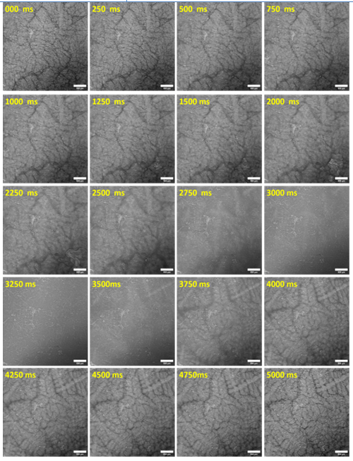
7. Dynamical imaging
Real time evolution of dendrite in Al-Cu alloy for observance of dendrite formation in the directional solidification experiment
Application Areas
Material Applications
Phase contrast imaging and Micro-tomography enables 3D Visualization of micro-structure such as voids, cracks, imperfections, material phases etc.
Structural features such as porosity, shape, size distribution of various phases, and geometrical parameters etc. can be quantified.
Mechanical and transport properties of material depends on its microstructure.
Structure properties correlations and modeling for optimization of material physical properties.
Study of effect of various sample environments on structural properties.
Biomedical Applications:
Diagnostic Imaging: Pre-clinical study and potential for clinical imaging - Mammography, Angiography, functional lung imaging etc.
Evolutionary research in species – comparison of organs at various stage of development.
Effects of environment, drug treatment and mutation over animals and plants.
Study of bio-materials and novel contrast agents.
Image guided radiation therapy.
Geology Applications
Rocks are used as construction material, resources of minerals, petrochemical reservoirs etc.
Micro-tomography study provides micro-structure which helps in quantification of its reservoir and transport properties.
Micro-tomography also quantifies mineral phase distribution.
Micro-structure of rocks is used to provide information about their origin and evolution of the rocks in the region.
1.
Deep learning assisted insights on porosity evolution of WAAM aluminium alloys. Babu A, M H, Agrawal AK, Shrawankar RC, Proceedings of the Institution of Mechanical Engineers, Part E: Journal of Process Mechanical Engineering. 2026;0(0). doi:10.1177/09544089261428355
1.
Micro-CT based internal morphological analysis of blowfly (Chrysomya megacephala): a potential tool to age blowfly pupae having forensic implications. Sharma, A., Agrawal, A.K., Singh, B. et al. Int J Trop Insect Sci (2025). https://doi.org/10.1007/s42690-025-01542-3
2.
Development of a novel single absorption grating based versatile multi-contrast imaging facility at the X-ray Imaging beamline, Indus-2. Balwant Singh, Ashish K. Agrawal, Yogesh Kashyap, Payal Singhai, Mayank Shukla; Rev. Sci. Instrum. 1 May 2025; 96 (5): 053705. https://doi.org/10.1063/5.0250945
3.
Quantitative analysis of reinforcement of nano-sized monetite particles on chitosan/gelatin 3D porous scaffolds using the micro-CT technique. Singh, Y.P., Dasgupta, S., Agrawal, A.K. et al. J Porous Mater (2025).https://doi.org/10.1007/s10934-025-01807-9
4.
Influence of chemical purification methodologies on the Bridgman growth of trans-stilbene (TSB) crystal, and feasibility studies for X-ray imaging and neutron/gamma discrimination applications. Pal, A., Kar, S., Debnath, C. et al. J Mater Sci: Mater Electron 36, 793 (2025).https://doi.org/10.1007/s10854-025-14845-5
5.
Optimization of selective laser sintering process parameters for phenolic thermoset composites using X-ray CT analysis, Jairam Raigar, Rajkumar Velu, Ashish Kumar Agrawal, Hadi Bakhshi, Materialia,Volume 42,2025, 102451, ISSN 2589-1529,https://doi.org/10.1016/j.mtla.2025.102451.
6.
A comparative study of correlation and moment analysis based phase retrieval techniques for unidirectional X-ray speckle interferometry.Kashyap, Y., Agrawal, A., Shukla, M. et al. J Opt (2025). https://doi.org/10.1007/s12596-025-02679-7
7.
A comparative study of sub-sampling methods in X-ray speckle interferometry based phase contrast imaging using synchrotron radiation source, Nuclear Instruments and Methods in Physics Research Section A:Yogesh Kashyap, Ashish Agrawal, Mayank Shukla, Hongchang Wang, Kawal Sahwney, Accelerators, Spectrometers, Detectors and Associated Equipment,Volume 1070, Part 1,2025, 170042, ISSN 0168-9002,https://doi.org/10.1016/j.nima.2024.170042.
8.
Development of Trans-stilbene/PMMA Polymer Composites and their Optical Characterization for Scintillation-based Imaging and Detector Applications; Chiranjit Debnath, Sujan Kar, Ashish K. Agrawal, Mayank Shukla, Sunil Verma; IEEE TRANSACTIONS ON NUCLEAR SCIENCE;DOI 10.1109/TNS.2024.3454716
1.
Enhancing microstructural integrity and mechanical performance of thick wire-arc directed energy deposited eutectic aluminum-silicon solid structure through continuous multi-pass friction stir processing; M. Hemachandra, Shivraman Thapliyal, Saravana Sundar, Adepu Kumar, J.P. Oliveira, Ashish K. Agrawal; Materials Science & Engineering A Volume 914, 2024,147155,ISSN 0921-5093 https://doi.org/10.1016/j.msea.2024.147155
2.
Damage kinetics and compression behavior of Al alloy foam using in situ SRμCT. Agrawal, A.K., Singhai, P., Singh, B. Kashyap Y.S. Shukla M. J Mater Sci (2024). https://doi.org/10.1007/s10853-024-09580-x
3.
Effect of Cu/Li Ratio on Porosity and Microstructural Evolution of Gravity and Squeeze-Cast Al–Cu–Li Alloys. Manojkumar, S., Agarwal, A.K., Roy, T. Mehta K.K..Metall Mater Trans B 55, 1117–1133 (2024).https://doi.org/10.1007/s11663-024-03028-y
4.
Quantifying pore characteristics in polymer glass–ceramics composite scaffolds using micro-tomography. Thomas, A., Agarwal, A.K., Kashyap, Y.S. Pravin Kumar I, Bera Japas. Journal of Materials Research (2024).https://doi.org/10.1557/s43578-024-01307-7
Study of Hot Deformation Behavior of EN25 Steel in the Presence of Non-metallic Inclusions. Singh, V., Roy, G.G., Srirangam, P. Chakraborty D. Agrawal A.K. J. of Materi Eng and Perform (2024). https://doi.org/10.1007/s11665-024-09247-3
7.
Quantitative phase contrast X-ray tomography of aluminium metal matrix composite, Ashish K. Agrawal, Chiradeep Gupta, Balwant Singh, Yogesh Kashyap, Mayank Shukla,Applied Radiation and Isotopes, Volume 204, 2024, 111149, ISSN 0969-8043, https://doi.org/10.1016/j.apradiso.2023.111149.
8.
Nano-fluorcanasite-fluorapatite Reinforced Poly-ε-caprolactone Based Biomimetic Scaffold: A Synergistic Approach Towards Generation of Conducive Environment for Cell Survival. Kumawat, V.S., Saini, R.K., Agrawal, A.K. et al. J Polym Environ 32, 411–429 (2024).https://doi.org/10.1007/s10924-023-02977-w
1.
Structural and characterization assessment of clay ceramic water filter materials from locations near the Thar Desert in India Sunil Duhan, S Gupta,Ashish Kumar Agrawal,A K Plappally;
Desalination and Water Treatment 309 (2023):236-248https://doi.org/10.1080/10.5004/dwt.2023.29879
2.
Study of thorium-induced micro-structural changes in mice femoral bone using SR-µCT. Agrawal, A.K., Yadav, R., Singh, B. Ali Manzoor , Kumar Amit , Kashyap Y.S. Pandey B. N.
Toxicol. Environ. Health Sci. (2023).https://doi.org/10.1007/s13530-023-00191-8
3.
Pore anisotropy in shale and its dependence on thermal maturity and organic carbon content: A scanning SAXS study, Jitendra Bahadur, Debanjan Chandra, Avik Das, Vikram Vishal, Ashish Kumar Agrawal, Debasis Sen, International Journal of Coal Geology,Volume 273, 2023,104268,ISSN 0166-5162,https://doi.org/10.1016/j.coal.2023.104268
1.
Effect of monetite reinforced into the chitosan-based lyophilized 3D scaffolds on physicochemical, mechanical, and osteogenic properties, International Journal of Polymeric Materials and Polymeric Biomaterials, Yogendra Pratap Singh, Rakesh Bhaskar, Ashish Kumar Agrawal & Sudip Dasgupta (2022) Effect of monetite reinforced into the chitosan-based lyophilized 3D scaffolds on physicochemical, mechanical, and osteogenic properties, International Journal of Polymeric Materials and Polymeric Biomaterials, 2022
https://doi.org/10.1080/00914037.2022.2090358
2.
Surfactant Assisted In Situ Synthesis of Nanofibrillated Cellulose/Polymethylsilsesquioxane Aerogel for Tuning Its Thermal Performance Pragya Gupta, Manoj Sathwane, Monika Chhajed, Chhavi Verma, Yves Grohens, Bastien Seantier, Ashish K. Agrawal, Pradip K. Maji Macromolecular Rapid Communications 2022 https://doi.org/10.1002/marc.202200628
3.
Bioactive fluorcanasite reinforced magnesium alloy-based porous bio-nanocomposite scaffolds with tunable mechanical properties Adithya Garimella, Ramya M, Subrata Bandhu Ghosh, Sanchita Bandyopadhyay-Ghosh, Ashish Kumar Agrawal; 2022 Journal of Biomedical Materials Research Part B: Applied Biomaterials https://doi.org/10.1002/jbm.b.35166
4.
Nano-scale physio-chemical attributes and their impact on pore heterogeneity in shale; Debanjan Chandra, Vikram Vishal, Jitendra Bahadur, Ashish Kumar Agrawal, Avik Das, Bodhisatwa Hazra, Debasis Sen,
Fuel, 314, 123070 (2022),
5.
Digital light processing mediated 3D printing of biocomposite bone scaffolds: Physico‐chemical interactions and in‐vitro biocompatibility; Abhijit Vyas,Subrata Bandhu Ghosh,Sanchita Bandyopadhyay-Ghosh,Ashish Kumar Agrawal,Deepak Khare,Ashutosh Kumar Dubey;
https://doi.org/10.1002/pc.26609
6.
Effect of graphene addition on thermal behavior of 3D printed graphene/AlSi10Mg composite, Jitendar Kumar Tiwari, Ajay Mandal, N. Sathish, Surender Kumar, Mohammed Ashiq, M. Nagini, R.K. Sharma, A.K. Agrawal, P. Rajput, A.K. Srivastava, Journal of Alloys and Compounds,Volume 890,2022,161725,
1.
The white beam station at imaging beamline BL-4, Indus-2; Agrawal, A. K., Singh, B., Singhai, P., Kashyap, Y. & Shukla, M. . J.
Synchrotron Rad. 28, 1639-1648 (2021).
2.
Monetite addition into gelatin based freeze-dried scaffolds for improved mechanical and osteogenic properties; Yogendra Pratap Singh, Sudip Dasgupta, Rakesh Bhaskar, Ashish Kumar Agrawal;
Biomed. Mater. 16, 065030 (2021)
3.
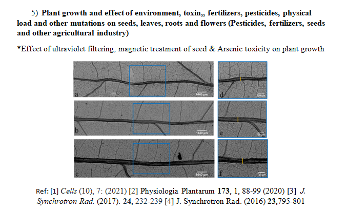
"Use of Synchrotron Phase-Sensitive Imaging for the Investigation of Magnetopriming and Solar UV-Exclusion Impact on Soybean (Glycine max) Leaves", Fatima, Anis, Sunita Kataria, Ashish K. Agrawal, Balwant Singh, Yogesh Kashyap, Meeta Jain, Marian Brestic, Suleyman I. Allakhverdiev, and Anshu Rastogi;
Cells 10, 7: 1725 (2021).
4.
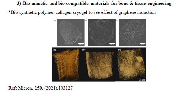
X-ray tomography analysis reveals the influence of graphene on porous morphology of collagen cryogels; Gopal Agarwal, Ashish Kumar Agrawal, Anis Fatima, Akshay Srivastava;
Micron, 150,103127,(2021).
Development of In-situ X-ray imaging and μCT facility under load conditions at imaging beamline Indus-2, Ashish K. Agarwal, Payal Singhai, B. Singh, Yogesh Kashyap and Mayank Shukla,
BARC News-Letter,16-20(374)
7.
Bio-extract amalgamated sodium alginate-cellulose nanofibres based 3Dsponges with interpenetrating BioPU coating as potential wound care scaffolds; Chandravati Yadav, Monika Chhajed, Priyanka Choudhury, Ram Prasad Sahu, Amit Patel, Saurabh Chawla, Luna Goswami, Chandan Goswami, Xinping Li, Ashish K. Agrawal, Arun Saini, Pradip K. Maji;
Materials Science & Engineering C 118 111348 (2021)
1.
Synchrotron radiation based micro computed tomographic analysis of dentinal microcracks using rotary and reciprocating file systems: An in vitro study; Harikumar Vemisetty, N Tulasi Priya, Balwant Singh, Pooja Yenubary, Ashish Kumar Agarwal, Jayaprada Reddy, Surakanti;
J Conserv Dent ;23:309 13 (2020)
2.
Correlationship of Drug-Polymer Miscibility, Molecular Relaxation and Phase Behavior of Dipyridamole Amorphous Solid Dispersions, Jagadish Sharma, Balwant Singh, Ashish Kumar Agrawal, Arvind K. Bansal,
Journal of Pharmaceutical Sciences, 0022-3549,(2020)
3.
Magneto priming effects on arsenic stress induced morphological and physiological variations in soybean involving synchrotron imaging; Anees Fatima; Sunita Kataria; Rajkumar Prajapati, ; Meeta jain; Ashish Agarwal; Balwant Singh; Y kashyap; Durgesh Kumar Tripathi; Vijay Pratap Singh; Rekha Gadre; ;
Physiologia Plantarum (2020);
4.
Tailor-made design, fabrication and validation of SrO doped nanostructured ZTA ceramic femoral head acetabular socket liner assembly, Shaik Akbar Basha, Ashish Kumar Agrawal, Debasish Sarkar,
Journal of the Mechanical Behavior of Biomedical Materials,,104178, (2020)
5.
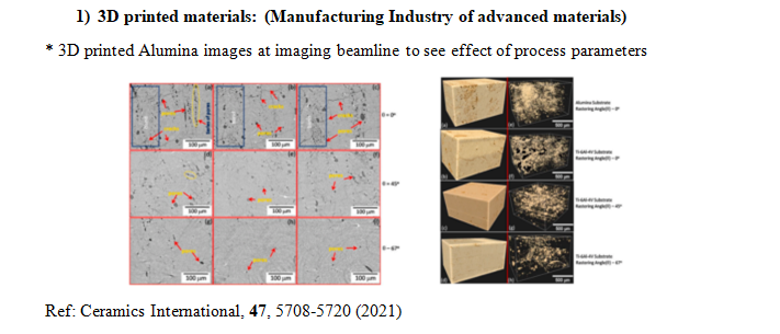
Experimental investigation on Laser Directed Energy Deposition based additive manufacturing of Al2O3 bulk structures, Govind Kumar Mishra, Christ Prakash Paul, Arun Kumar Rai, Ashish Kumar Agrawal, Sanjay Kumar Rai, Kushvinder Singh Bindra,
Ceramics International,2020, ISSN 0272-8842, https://doi.org/10.1016/j.ceramint.2020.10.157.
6.
Fabrication of guar gum-gelatin scaffold for soft tissue engineering, Abhishek Indurkar, Prachi Bangde, Manish Gore, Ashish K. Agrawal, Ratnesh Jain, Prajakta Dandekar,
Carbohydrate Polymer Technologies and Applications, 1, , 100006, (2020)
7.
Evaluation of Influence of Finish line Design on Marginal Discrepancy of All-ceramics Lithium disilicate Crown restorations using μ-CT; Yajvinder, Vishal Gulati, Ashish Agrawal, Balwant Singh,
Materials Science and Engineering 802 012003 (2020)
8.
Demineralization of tooth enamel following radiation therapy; An in vitro microstructure and micro-hardness analysis; Jagadish Kudkuli, Ashish Agrawal,Om Prakash Gurjar, Sunil Dutt Sharma, P. D. Rekha, Muhammed A. P. Manzoor, Balwant Singh, B. S. Rao, Riaz Abdulla,
J Can Res Ther May (2020)
9.
Preparation and application of silica nanoparticles-Ocimum basilicum seeds bio-hybrid for the efficient immobilization of invertase enzyme; Archana Mishra, Jose Savio Melo*, Ashish K. Agrawal, Yogesh Kashyap and Debasis Sen;
Colloids and Surfaces B: Biointerfaces 188 110796 (2020)
10.
Characterization Of Micro And Meso Porosity In Portland Cement At Elevated Temperatures. P. Harsha Praneeth, Tezeswi P. Tadepalli, Ashish K. Agrawal:
Magazine of Concrete Research 72 (6), 304-313; DOI:10.1680/jmacr.18.00321 (2020)
11.
Thermal Behavior of PPC and OPC-53 When Exposed to Extreme Temperatures. Harsha Praneeth Pavani, T. P. Tezeswi, Ashish Kumar Agrawal:
Advances in Cement Research 32 (8), 358-370; DOI:10.1680/jadcr.18.00066 (2020)
12.
Investigation of porosity, microstructure and mechanical properties of additively manufactured graphene reinforced AlSi10Mg composite; J. K. Tiwari, Ajay Mandal, N.Sathish, A.K.Agrawal, A.K.Srivastava,
Additive Manufacturing, 33, 101095 (2020)
1.
Study of gamma irradiation induced micro-structural variations in flame retardant polyurethane foam using synchrotron X-ray micro-CT; Ashish Agrawal, Balwant Singh, Y. S. Kashyap, Mayank Shukla, S.C. Gadkari;
J. Synchrotron Rad. 26 (5) 2019 1797-1807 (2019)
2.
Preparation and characterization of glass–ceramic reinforced alginate scaffolds for bone tissue engineering Ashley Thomas, Eldin Johnson, Ashish K. Agrawal, Japes bera;
Advanced Engineering materials; 34, (22-28) , pp. 3798-3809 (2019)
3.
Esterified superhydrophobic nano-fibrillated cellulose based aerogel for oil spill treatment; Monika Chhajed, Chandravati Yadav, Ashish K. Agrawal, Pradip K. Maji;
Carbohydrate Polymers 226 115286 (2019)
4.
Morphological characteristics & microstructure of kidney stones using synchrotron radiation μ-CT reveal the mechanism of crystal growth and aggregation in mixed stones. M. A. P. Manzoor, Ashish K. Agrawal, Balwant Singh, M. Mujeeburahiman, Punchappady Devasya Rekha PLoS ONE 14(3):
e0214003 (2019)
5.
Establishing Correlations among Pore Structure, Surface Roughness, Compressive Strength and Fracture Toughness of Ceramic Water Filters Local to Rajasthan, India, A. Kaurwar, A.K Agarwal, B. Singh, S. Gupta, R. Satankar, J. M. Oommen, L. Dave, M. Sharif, A.B.O Soboyejo, A. Plappally
Desalination and Water Treatment 157 332–341 (2019)
6.
Investigations on high strength macroporous Silicon Oxycarbide ceramics with the aid of synchrotron based µ-Tomography; Abhisek Choudhary, Ashish K. Agrawal, Balwant Singh, Swadesh Kumar Pratihar, Shantanu K Behera;
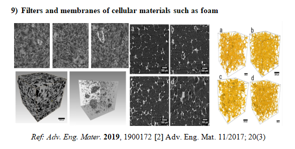
Adv. Eng. Mater. 1900172 (2019)
7.
Morphological and elemental mapping of Gallstones using Synchrotron Micro tomography (SR- µCT) and Synchrotron X-ray Fluorescence Spectroscopy (SRXRF); B. Mohana , Jayanthi Venkataraman , Ramya JR , Mayank Jain , Balwant Singh , K. Thanigai Arul, Vaithiswaran V , Saravanan M N, Tiwari M K, Agarwal A K , S. Narayana Kalkuraa
JGH Open: 1–7 (2019) doi:10.1002/jgh3.12171
8.
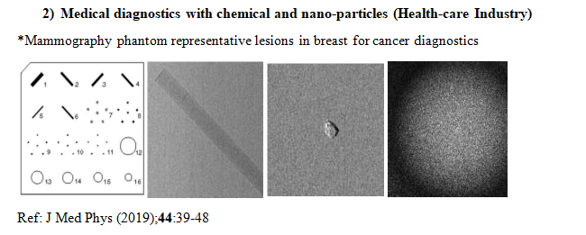
Phantom based feasibility studies on phase-contrast mammography at Indian synchrotron facility Indus-2; Reena Sharma, S D Sharma, P S Sarkar, B Singh, Dr. A K Agrawal, D Datta;
J Med Phys ;44:39-48 (2019)
9.
3D spatial distribution of ore mineral phases using high-resolution synchrotron Micro-Computed Tomography (µCT) combined with Optical Microscopy. A. Fatima, A. S. Venkatesh, R. Mukherjee, A. K. Agrawal, B.Singh, P. S. Sarkar, Y. Kashyap, T. Shripathi
Applied Radiation and Isotopes 148, 49–59 (2019)
10.
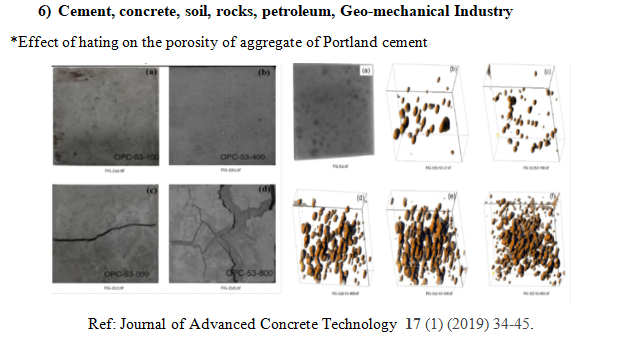
Estimation of Porosity and Pore Distribution in Hydrated Portland Cement at Elevated Temperatures using Synchrotron Micro Tomography; Harsha Praneeth, Tezeswi Tadepalli, Ashish Kumar Agrawal:
Journal of Advanced Concrete Technology 17 (1) 34-45,(2019)
1.
Morphological and micro-tomographic study on evolution of struvite in synthetic urine infected with bacteria and investigation of its pathological biomineralization. Muhammed A. P. Manzoor, Balwant Singh, Ashish K. Agrawal, Ananthapadmanabha Bhagwath Arun, M. Mujeeburahiman, Punchappady Devasya Rekha:
PLoS ONE ; 13(8):e0202306., (2018)
2.
Low density and high strength nano-fibrillated cellulose aerogel for thermal insulation application. Pragya Gupta, Balwant Singh, Ashish K. Agrawal, Pradip K. Maji:
Materials and Design ; 158; 224–236 (2018)
3.
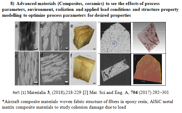
True damage state and cracking in highly deformed metal matrix composites revealed by synchrotron microtomography. Chiradeep Gupta, Ashish K Agarwal, Balwant Singh, S.C. Gadkari, Krishnan Madangopal:
Materialia 3, 218-229 (2018)
4.
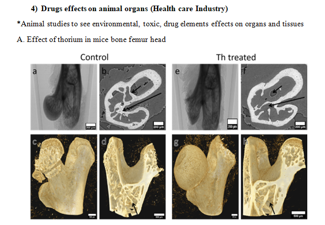
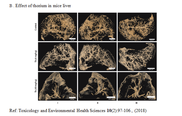
Thorium-induced Anatomical and Histopathological Changes in Liver of Swiss Mice. Rakhee Yadav, Ashish K. Agrawal, Manjoor Ali, Amit Kumar, Balwant Singh, Yogesh Kashyap, Amar Sinha, S. C. Gadkari, Badri N. Pandey:
Toxicology and Environmental Health Sciences ; 10(2):97-106.(2018)
1.
Macroporous SiOC Ceramics with Dense Struts by Positive Sponge Replication Technique; Abhisek Choudhary, Swadesh K. Pratihar, Ashish K. Agrawal, Shantanu K. Behera:
Advanced Engineering Materials ; 20(3).(2017), DOI:10.1002/adem.201700586
2.
Surface Plasmon Band Tailoring of Plasmonic Nanostructure under the Effect of Water Radiolysis by Synchrotron Radiations; Amardeep Bharti, Ashish K Agrawal, Balwant Singh, Sanjeev Gautam, Navdeep Goyal,
J. Synchrotron Rad. 24 (2017)
3.
Study of damage development near inhomogeneous compression regions of hot deformed SiCP/A6061 composite; Materials Science & Engineering A; Chiradeep Gupta, Ashish K.Agarwal Balwant Singh Partha S.Sarkar S.C.Gadkari AmarSinha Dinesh Srivastava Gautam K.Dey;
704292–301, (2017)
4.
Synchrotron based X-ray micro-imaging facility for bio-medical research; Ashish Kumar Agrawal, Balwant Singh, Y.S. Kashyap, Mayank Shukla, S.C. Gadkari;
J Radiation & Cancer Res ;8:153-9 (2017)
5.
Synchrotron based phase contrast tomography of hypercholesteromic rat liver A. Fatima, A. K. Agrawal, B. Singh, D. Bhatnagar, H. Sharma, T. Shripathi and Y. Kashyap.;
International Journal of Medical Research & Health Sciences, , 6(5): 115-125 (2017)
6.
Synchrotron-based phase-sensitive imaging of leaves grown from magneto-primed seeds of soybean; A. Fatima, S. Kataria, L. Baghel, K. N. Guruprasad, A. K. Agrawal, B. Singh, P. S. Sarkar, T. Shripathi, Y. Kashyap;
J. Synchrotron Rad. . 24, 232-239 (2017)
1.
Monochromatic X-ray induced novel synthesis of plasmonic nano-structures for photovoltaic application; Amardeep Bharti, Richa Bhardwaj, Ashish K. Agrawal, Navdeep Goyal, Sanjeev Gautam;
Nature Scientific Reports 6 (22394) (2016).
2.
Synchrotron hard X-ray micro-imaging of leaf venation in soybean (Glycine max) after exclusion of solar UV (280-400nm); A. Fatima, S. Kataria, K.N. Guruprasad, A. K. Agrawal, B. Singh, P. S. Sarkar, T. Shripathi, Y. Kashyap and A. Sinha,
J. Synchrotron Rad. 23,795-801 (2016)
3.
Non-destructive evaluation of teeth restored with different resins using synchrotron based micro-imaging; A. Fatima, V.K. Kulkarni, N.R. Banda, A.K. Agrawal, B. Singh, P. S. Sarkar, S. Tripathi, T. Shripathi, Y. Kashyap, A. Sinha;
Journal of X-ray Science and technology 24 119–132 (2016).
1.
Design, development and first experiments on the X-ray Imaging Beamline at Indus-2 synchrotron source RRCAT, India, A. K. Agrawal, B. Singh, Y.S. Kashyap, M. Shukla, P. S. Sarkar, A. Sinha;
J. Synchrotron Rad. 22, 1531–1539 (2015).